The Cole Neurocognition Lab

A cognitive, computational, and network neuroscience laboratory located at the Center for Molecular and Behavioral Neuroscience (CMBN) at Rutgers University


Research Projects


In our research we seek connectivity-based explanations of neurocognitive phenomena, especially those related to learning and goal-directed cognition.

We achieve this using a variety of techniques, applying network science, computational modeling, and machine learning approaches to data collected from the human brain (with fMRI, MEG, EEG, diffusion MRI, and behavioral measures) and neural network simulations.

Much of this work involves understanding the role of brain connectivity in producing the computations apparent in task-driven brain activity patterns and behavior. This facilitates theoretical understanding of cognitive processes as they are generated by brain network interactions, providing insights into both natural and artificial intelligence.

Our ultimate goal is to utilize brain connectivity research to advance fundamental understanding of the human brain, driving applications that enhance the human condition – especially via novel treatments for brain diseases such as major depression, Alzheimer's disease, and schizophrenia.


Neurocognitive Basis of Cognitive Control

Cognitive control consists of the processes underlying goal-directed cognition – processes important for everyday life and disrupted in a variety of mental illnesses. Cognitive control is relevant to most thoughts and behaviors, such as learning, emotion regulation, attention, problem solving, intelligence, and memory.
Network Mechanisms of Cognitive Control
  • Ito T, Yang GB, Laurent P, Schultz DH, Cole MW (2022). "Constructing neural network models from brain data reveals representational transformations linked to adaptive behavior". Nature Communications. 13, 673. doi:10.1038/s41467-022-28323-7 Code On Twitter
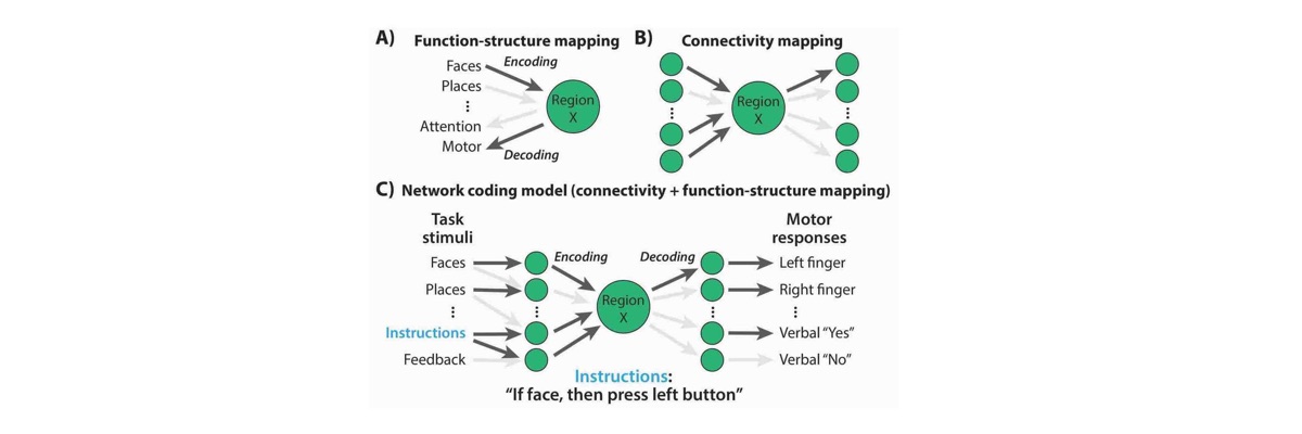
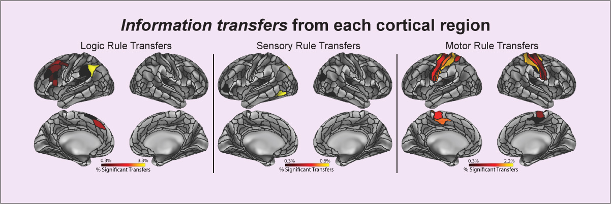
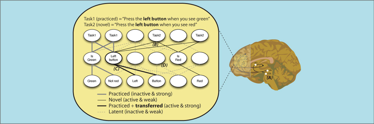
    The human ability to adaptively implement a wide variety of tasks is thought to emerge from the dynamic transformation of cognitive information. We hypothesized that these transformations are implemented via conjunctive activations in “conjunction hubs”—brain regions that selectively integrate sensory, cognitive, and motor activations. We used recent advances in using functional connectivity to map the flow of activity between brain regions to construct a task-performing neural network model from fMRI data during a cognitive control task. We verified the importance of conjunction hubs in cognitive computations by simulating neural activity flow over this empirically-estimated functional connectivity model. These empirically-specified simulations produced above-chance task performance (motor responses) by integrating sensory and task rule activations in conjunction hubs. These findings reveal the role of conjunction hubs in supporting flexible cognitive computations, while demonstrating the feasibility of using empirically-estimated neural network models to gain insight into cognitive computations in the human brain.
  • Cocuzza CV, Ito T, Schultz D, Bassett DS, Cole MW (2020). "Flexible coordinator and switcher hubs for adaptive task control". Journal of Neuroscience. 40(36):6949–6968. doi:10.1523/JNEUROSCI.2559-19.2020 On Twitter On Facebook Blog Post Code
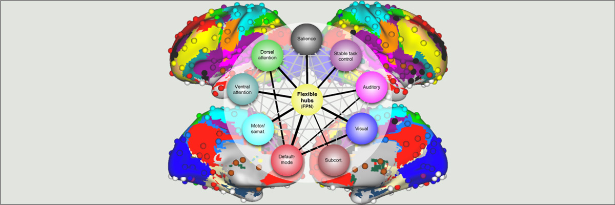
    Functional connectivity studies have identified at least two large-scale neural systems that constitute cognitive control networks – the frontoparietal network (FPN) and cingulo-opercular network (CON). Control networks are thought to support goal-directed cognition and behavior. It was previously shown that the FPN flexibly shifts its global connectivity pattern according to task goal, consistent with a “flexible hub” mechanism for cognitive control. Our aim was to build on this finding to develop a functional cartography (a multi-metric profile) of control networks in terms of dynamic network properties. We quantified network properties in (male and female) humans using a high-control-demand cognitive paradigm involving switching among 64 task sets. We hypothesized that cognitive control is enacted by the FPN and CON via distinct but complementary roles reflected in network dynamics. Consistent with a flexible “coordinator” mechanism, FPN connections were globally diverse across tasks, while maintaining within-network connectivity to aid cross-region coordination. Consistent with a flexible “switcher” mechanism, CON regions switched to other networks in a task-dependent manner, driven primarily by reduced within-network connections to other CON regions. This pattern of results suggests FPN acts as a dynamic, global coordinator of goal-relevant information, while CON transiently disbands to lend processing resources to other goal-relevant networks. This cartography of network dynamics reveals a dissociation between two prominent cognitive control networks, suggesting complementary mechanisms underlying goal-directed cognition.
  • Cole M.W., Reynolds J.R., Power J.D., Repovs G., Anticevic A., Braver T.S. (2013). "Multi-task connectivity reveals flexible hubs for adaptive task control". Nature Neuroscience; 2013 Sep;16(9):1348-55. doi:10.1038/nn.3470. More info
    Extensive evidence suggests the human ability to adaptively implement a wide variety of tasks is preferentially due to the operation of a fronto-parietal brain network. We hypothesized that this network’s adaptability is made possible by ‘flexible hubs’ – brain regions that rapidly update their pattern of global functional connectivity according to task demands. We utilized recent advances in characterizing brain network organization and dynamics to identify mechanisms consistent with the flexible hub theory. We found that the fronto-parietal network’s brain-wide functional connectivity pattern shifted more than other networks’ across a variety of task states, and that these connectivity patterns could be used to identify the current task. Further, these patterns were consistent across practiced and novel tasks, suggesting reuse of flexible hub connectivity patterns facilitates adaptive (novel) task performance. Together, these findings support a central role for fronto-parietal flexible hubs in cognitive control and adaptive implementation of task demands generally.
  • Cole M.W., Yarkoni T., Repovs G., Anticevic A., Braver T.S. (2012). "Global Connectivity of Prefrontal Cortex Predicts Cognitive Control and Intelligence". Journal of Neuroscience. 32(26): 8988-8999; doi: 10.1523/JNEUROSCI.0536-12.2012
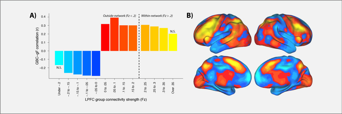
    Control of thought and behavior is fundamental to human intelligence. Evidence suggests a frontoparietal brain network implements such cognitive control across diverse contexts. We identify a mechanism — global connectivity — by which components of this network might coordinate control of other networks. A lateral prefrontal cortex (LPFC) region’s activity was found to predict performance in a high control demand working memory task and also to exhibit high global connectivity. Critically, global connectivity in this LPFC region, involving connections both within and outside the frontoparietal network, showed a highly selective relationship with individual differences in fluid intelligence. These findings suggest LPFC is a global hub with a brainwide influence that facilitates the ability to implement control processes central to human intelligence.
  • Cole M.W., Schneider W. (2007). “The cognitive control network: Integrated cortical regions with dissociable functions”, NeuroImage 37(1): 343-360. doi: 10.1016/j.neuroimage.2007.03.071
    Consensus across hundreds of published studies indicates that the same regions are involved in many forms of cognitive control. Using functional magnetic resonance imaging (fMRI), we found that these coactive regions form a functionally connected cognitive control network (CCN). Network status was identified by convergent methods, including: high interregional correlations during rest and task performance, consistently higher correlations within the CCN than the rest of cortex, co-activation in a visual search task, and mutual sensitivity to decision difficulty. Regions within the CCN include anterior cingulate cortex / pre-supplementary motor area (ACC/pSMA), dorsolateral prefrontal cortex (DLPFC), inferior frontal junction (IFJ), anterior insular cortex (AIC), dorsal pre-motor cortex (dPMC), and posterior parietal cortex (PPC). We used a novel visual line search task which included periods when the probe stimuli were occluded but subjects had to maintain and update working memory in preparation for the sudden appearance of a probe stimulus. The six CCN regions operated as a tightly coupled network during the ‘non-occluded’ portions of this task, with all regions responding to probe events. In contrast, the network was differentiated during occluded search. DLPFC, not ACC/pSMA, was involved in target memory maintenance when probes were absent, while both regions became active in preparation for difficult probes at the end of each occluded period. This approach illustrates one way in which a neuronal network can be identified, its high functional connectivity established, and its components dissociated in order to better understand the interactive and specialized internal mechanisms of that network.
Rapid Instructed Task Learning (RITL; "rittle")
  • Cole M.W., Ito T., Braver T.S. (2016). "The behavioral relevance of task information in human prefrontal cortex". Cerebral Cortex. doi: 10.1093/cercor/bhv072
    Human lateral prefrontal cortex (LPFC) is thought to play a critical role in enabling cognitive flexibility, particularly when performing novel tasks. However, it remains to be established whether LPFC representation of task-relevant information in such situations actually contributes to successful performance. We utilized pattern classification analyses of functional MRI activity to identify novelty-sensitive brain regions as participants rapidly switched between performance of 64 complex tasks, 60 of which were novel. In three of these novelty-sensitive regions – located within distinct areas of left anterior LPFC – trial-evoked activity patterns discriminated correct from error trials. Further, these regions also contained information regarding the task-relevant decision rule, but only for successfully performed trials. This suggests that left anterior LPFC may be particularly important for representing task information that contributes to the cognitive flexibility needed to perform successfully in novel task situations.
  • Cole M.W., Laurent P., Stocco A. (2013). "Rapid instructed task learning: A new window into the human brain’s unique capacity for flexible cognitive control". Cognitive, Affective, & Behavioral Neuroscience. 13(1): 1-22; doi:10.3758/s13415-012-0125-7
    The human ability to flexibly adapt to novel circumstances is extraordinary. Perhaps the most illustrative, yet underappreciated, form of this cognitive flexibility is rapid instructed task learning (RITL) – the ability to rapidly reconfigure our minds to perform new tasks from instructions. This ability is important for everyday life (e.g., learning to use new technologies) and is used to instruct participants in nearly every study of human cognition. We review the development of RITL as a circumscribed domain of cognitive neuroscience investigation, culminating in recent demonstrations that RITL is implemented via brain circuits centered on lateral prefrontal cortex. We then build on this and the recent discovery of compositional representations within lateral prefrontal cortex to develop an integrative theory of cognitive flexibility and cognitive control that identifies mechanisms that may enable RITL within the human brain. The insights gained from this new theoretical account have important implications for further developments and applications of RITL research.
  • Cole M.W., Bagic A., Kass R., Schneider W. (2010). "Prefrontal Dynamics Underlying Rapid Instructed Task Learning Reverse With Practice". Journal of Neuroscience 30(42):14245-14254. doi: 10.1523/JNEUROSCI.1662-10.2010.
    The ability to rapidly reconfigure our minds to perform novel tasks is important for adapting to an ever-changing world, yet little is understood about its basis in the brain. Further, it is unclear how this kind of task preparation changes with practice. Previous research suggests that prefrontal cortex (PFC) is essential when preparing to perform either novel or practiced tasks. Building upon recent evidence that PFC is organized in an anterior-to-posterior hierarchy, we postulated that novel and practiced task preparation would differentiate hierarchically distinct regions within PFC across time. Specifically, we hypothesized and confirmed using functional MRI and magnetoencephalography with humans that novel task preparation is a bottom-up process that involves lower-level rule representations in dorsolateral PFC (DLPFC) prior to a higher-level rule-integrating task representation in anterior PFC (aPFC). In contrast, we identified a complete reversal of this activity pattern during practiced task preparation. Specifically, we found that practiced task preparation is a top-down process that involves a higher-level rule-integrating task representation (recalled from long-term memory) in aPFC prior to lower-level rule representations in DLPFC. These findings reveal two distinct yet highly inter-related mechanisms for task preparation, one involving task set formation from instructions during rapid instructed task learning and the other involving task set retrieval from long-term memory to facilitate familiar task performance. These two mechanisms demonstrate the exceptional flexibility of human PFC as it rapidly reconfigures cognitive brain networks to implement a wide variety of possible tasks.
Dynamic Brain Network Organization

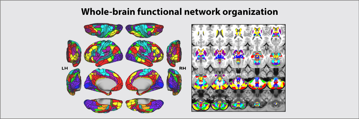
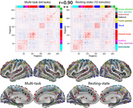
Behavior emerges from the brain's network architecture and the dynamic symphonies that play out on that architecture. We use functional and structural connectivity approaches to characterize properties of large-scale human brain networks, both at rest and during a wide variety of tasks. We apply existing and novel methods from network science and related areas to achieve these ends.
Select Publications:
  • Sanchez-Romero R, Ito T, Mill RD, Hanson SJ, Cole MW (2023). "Causally informed activity flow models provide mechanistic insight into network-generated cognitive activations". NeuroImage. 278:120300. doi:10.1016/j.neuroimage.2023.120300 On Twitter
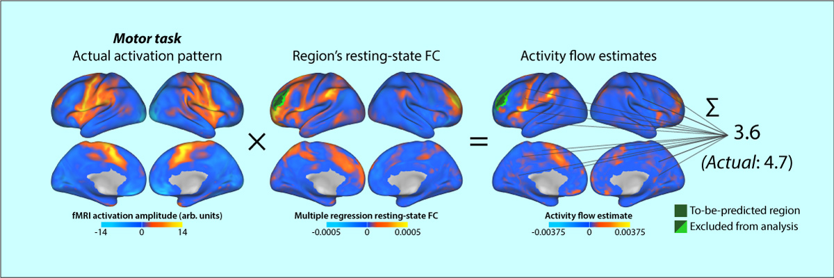
    Brain activity flow models estimate the movement of task-evoked activity over brain connections to help explain network-generated task functionality. Activity flow models have been shown to accurately generate task-evoked brain activations across a wide variety of brain regions and task conditions. However, these models have had limited explanatory power, given known issues with causal interpretations of the standard functional connectivity measures used to parameterize activity flow models. We show here that functional/effective connectivity (FC) measures grounded in causal principles facilitate mechanistic interpretation of activity flow models. We progress from simple to complex FC measures, with each adding algorithmic details reflecting causal principles. This reflects many neuroscientists’ preference for reduced FC measure complexity (to minimize assumptions, minimize compute time, and fully comprehend and easily communicate methodological details), which potentially trades off with causal validity. We start with Pearson correlation (the current field standard) to remain maximally relevant to the field, estimating causal validity across a range of FC measures using simulations and empirical fMRI data. Finally, we apply causal-FC-based activity flow modeling to a dorsolateral prefrontal cortex region (DLPFC), demonstrating distributed causal network mechanisms contributing to its strong activation during a working memory task. Notably, this fully distributed model is able to account for DLPFC working memory effects traditionally thought to rely primarily on within-region (i.e., not distributed) recurrent processes. Together, these results reveal the promise of parameterizing activity flow models using causal FC methods to identify network mechanisms underlying cognitive computations in the human brain.
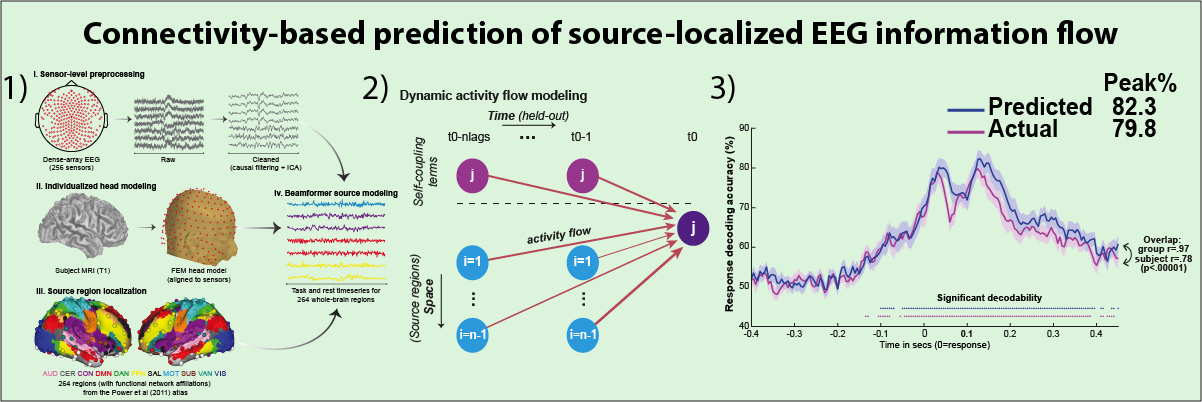
  • Mill RD, Hamilton JL, Winfield EC, Lalta N, Chen RH, Cole MW (2022). "Network modeling of dynamic brain interactions predicts emergence of neural information that supports human cognitive behavior". PLOS Biology. doi:10.1101/2021.01.26.428276 Commentary on paper Code On Twitter
    How cognitive task behavior is generated by brain network interactions is a central question in neuroscience. Answering this question calls for the development of novel analysis tools that can firstly capture neural signatures of task information with high spatial and temporal precision (the “where and when”) and then allow for empirical testing of alternative network models of brain function that link information to behavior (the “how”). We outline a novel network modeling approach suited to this purpose that is applied to noninvasive functional neuroimaging data in humans. We first dynamically decoded the spatiotemporal signatures of task information in the human brain by combining MRI-individualized source electroencephalography (EEG) with multivariate pattern analysis (MVPA). A newly developed network modeling approach—dynamic activity flow modeling—then simulated the flow of task-evoked activity over more causally interpretable (relative to standard functional connectivity [FC] approaches) resting-state functional connections (dynamic, lagged, direct, and directional). We demonstrate the utility of this modeling approach by applying it to elucidate network processes underlying sensory–motor information flow in the brain, revealing accurate predictions of empirical response information dynamics underlying behavior. Extending the model toward simulating network lesions suggested a role for the cognitive control networks (CCNs) as primary drivers of response information flow, transitioning from early dorsal attention network-dominated sensory-to-response transformation to later collaborative CCN engagement during response selection. These results demonstrate the utility of the dynamic activity flow modeling approach in identifying the generative network processes underlying neurocognitive phenomena.
  • Cole MW, Ito T, Bassett DS, & Schultz DH (2016). "Activity flow over resting-state networks shapes cognitive task activations". Nature Neuroscience. doi:10.1038/nn.4406
    Resting-state functional connectivity (FC) has helped reveal the intrinsic network organization of the human brain, yet its relevance to cognitive task activations has been unclear. Uncertainty remains despite evidence that resting-state FC patterns are highly similar to cognitive task activation patterns. Identifying the distributed processes that shape localized cognitive task activations may help reveal why resting-state FC is so strongly related to cognitive task activations. We found that estimating task-evoked activity flow (the spread of activation amplitudes) over resting-state FC networks allowed prediction of cognitive task activations in a large-scale neural network model. Applying this insight to empirical functional MRI data, we found that cognitive task activations can be predicted in held-out brain regions (and held-out individuals) via estimated activity flow over resting-state FC networks. This suggests that task-evoked activity flow over intrinsic networks is a large-scale mechanism explaining the relevance of resting-state FC to cognitive task activations.
  • Schultz DH & Cole MW (2016). "Higher Intelligence Is Associated with Less Task-Related Brain Network Reconfiguration". Journal of Neuroscience. doi:10.1523/JNEUROSCI.0358-16.2016
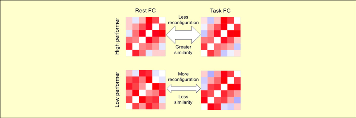
    The human brain is able to exceed modern computers on multiple computational demands (e.g., language, planning) using a small fraction of the energy. The mystery of how the brain can be so efficient is compounded by recent evidence that all brain regions are constantly active as they interact in so-called resting-state networks (RSNs). To investigate the brain's ability to process complex cognitive demands efficiently, we compared functional connectivity (FC) during rest and multiple highly distinct tasks. We found previously that RSNs are present during a wide variety of tasks and that tasks only minimally modify FC patterns throughout the brain. Here, we tested the hypothesis that, although subtle, these task-evoked FC updates from rest nonetheless contribute strongly to behavioral performance. One might expect that larger changes in FC reflect optimization of networks for the task at hand, improving behavioral performance. Alternatively, smaller changes in FC could reflect optimization for efficient (i.e., small) network updates, reducing processing demands to improve behavioral performance. We found across three task domains that high-performing individuals exhibited more efficient brain connectivity updates in the form of smaller changes in functional network architecture between rest and task. These smaller changes suggest that individuals with an optimized intrinsic network configuration for domain-general task performance experience more efficient network updates generally. Confirming this, network update efficiency correlated with general intelligence. The brain's reconfiguration efficiency therefore appears to be a key feature contributing to both its network dynamics and general cognitive ability.
  • Cole M.W., Bassett D.S., Power J.D., Braver T.S., Petersen S.E. (2014). "Intrinsic and task-evoked network architectures of the human brain". Neuron. 83(1), 238 – 251. doi:10.1016/j.neuron.2014.05.014
    Many functional network properties of the human brain have been identified during rest and task states, yet it remains unclear how the two relate. We identified a whole-brain network architecture present across dozens of task states that was highly similar to the resting-state network architecture. The most frequent functional connectivity strengths across tasks closely matched the strengths observed at rest, suggesting this is an “intrinsic”, standard architecture of functional brain organization. Further, a set of small but consistent changes common across tasks suggests the existence of a task-general network architecture distinguishing task states from rest. These results indicate the brain’s functional network architecture during task performance is shaped primarily by an intrinsic network architecture that is also present during rest, and secondarily by evoked task-general and task-specific network changes. This establishes a strong relationship between resting-state functional connectivity and task-evoked functional connectivity – areas of neuroscientific inquiry typically considered separately.

Network Disruptions Underlying Brain Disorders and Aging

We also investigate how the brain network properties identified in the first two research programs may explain symptoms of brain disorders, as well as effects of (healthy and unhealthy) aging. This works on two levels: First, cognitive control deficits are present across many brain/mental disorders and also emerge with aging. Second, we found that similar brain network disruptions are present across multiple brain disorders. Thus, discoveries in these areas may facilitate understanding of the deficits underlying brain disorders, possibly leading to treatments that apply across multiple disorders (and potentially aging as well).
Select Publications:
  • Hearne LJ, Mill RD, Keane BP, Repovs G, Anticevic A, Cole MW (2021). "Activity flow underlying abnormalities in brain activations and cognition in schizophrenia". Science Advances. 7(9) doi:10.1126/sciadv.abf2513 Code On Twitter
    Cognitive dysfunction is a core feature of many brain disorders such as schizophrenia (SZ), and has been linked to both aberrant brain functional connectivity (FC) and aberrant cognitive brain activations. We propose that aberrant network activity flow over FC pathways leads to altered cognitive activations that produce cognitive dysfunction in SZ. We tested this hypothesis using activity flow mapping – an approach that models the movement of task-related activity between brain regions as a function of FC. Using fMRI data from SZ individuals and healthy controls during a working memory task, we found that activity flow models accurately predict aberrant cognitive activations across multiple brain networks. Within the same framework, we simulated a connectivity-based clinical intervention, predicting specific treatments that normalized brain activations and behavior in independent patients. Our results suggest that dysfunctional task-evoked activity flow is a large-scale network mechanism contributing to the emergence of cognitive dysfunction in SZ.
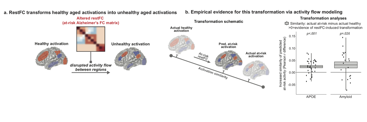
  • Mill RD, Gordon BA, Balota DA, Cole MW (2020). "Predicting dysfunctional age-related task activations from resting-state network alterations". NeuroImage. doi:10.1016/j.neuroimage.2020.117167 On Twitter Code
    Alzheimer’s disease (AD) is linked to changes in fMRI task activations and fMRI resting-state functional connectivity (restFC), which can emerge early in the timecourse of illness. Study of these fMRI correlates of unhealthy aging has been conducted in largely separate subfields. Taking inspiration from neural network simulations, we propose a unifying mechanism wherein restFC network alterations associated with Alzheimer’s disease disrupt the ability for activations to flow between brain regions, leading to aberrant task activations. We apply this activity flow modeling framework in a large sample of clinically unimpaired older adults, which was segregated into healthy (low-risk) and at-risk subgroups based on established imaging (positron emission tomography amyloid) and genetic (apolipoprotein) risk factors for AD. We identified healthy task activations in individuals at low risk for AD, and then by estimating activity flow using at-risk AD restFC data we were able to predict the altered at-risk AD task activations. Thus, modeling the flow of healthy activations over at-risk AD connectivity effectively transformed the healthy aged activations into unhealthy aged activations. These results provide evidence that activity flow over altered intrinsic functional connections may act as a mechanism underlying Alzheimer’s-related dysfunction, even in very early stages of the illness. Beyond these mechanistic insights linking restFC with cognitive task activations, this approach has potential clinical utility as it enables prediction of task activations and associated cognitive dysfunction in individuals without requiring them to perform in-scanner cognitive tasks.
  • Schultz DH, Ito T, Solomyak L, Chen RH, Mill RD, Anticevic A, Cole MW (2019). "Global connectivity of the frontoparietal cognitive control network is related to depression symptoms in the general population". Network Neuroscience. 3:1:107-123. doi:10.1162/NETN_a_00056 On Twitter On Facebook Blog Post
    We all vary in our mental health, even among people not meeting diagnostic criteria for mental illness. Understanding this individual variability may reveal factors driving the risk for mental illness, as well as factors driving sub-clinical problems that still adversely affect quality of life. To better understand the large-scale brain network mechanisms underlying this variability we examined the relationship between mental health symptoms and resting-state functional connectivity patterns in cognitive control systems. One such system is the frontoparietal cognitive control network (FPN). Changes in FPN connectivity may impact mental health by disrupting the ability to regulate symptoms in a goal-directed manner. Here we test the hypothesis that FPN dysconnectivity relates to mental health symptoms even among individuals who do not meet formal diagnostic criteria but may exhibit meaningful symptom variation. We found that depression symptoms severity negatively correlated with between-network global connectivity (BGC) of the FPN. This suggests that decreased connectivity between the FPN and the rest of the brain is related to increased depression symptoms in the general population. These findings complement previous clinical studies to support the hypothesis that global FPN connectivity contributes to the regulation of mental health symptoms across both health and disease.
  • Spronk M, Keane BP, Ito T, Kulkarni K, Ji JL, Anticevic A, Cole MW (2021). "A whole-brain and cross-diagnostic perspective on functional brain network dysfunction". Cerebral Cortex. doi:10.1093/cercor/bhaa242 On Twitter
    A wide variety of mental disorders have been associated with resting-state functional network alterations, which are thought to contribute to the cognitive changes underlying mental illness. These observations have seemed to support various theories postulating large-scale disruptions of brain systems in mental illness. However, existing approaches isolate differences in network organization without putting those differences in broad, whole-brain perspective. Using a graph distance measure - connectome-wide correlation - we found that whole-brain resting-state functional network organization in humans is highly similar across a variety of mental diseases and healthy controls. This similarity was observed across autism spectrum disorder, attention-deficit hyperactivity disorder, and schizophrenia. Nonetheless, subtle differences in network graph distance were predictive of diagnosis, suggesting that while functional connectomes differ little across health and disease those differences are informative. Such small network alterations may reflect the fact that most psychiatric patients maintain overall cognitive abilities similar to those of healthy individuals (relative to, e.g., the most severe schizophrenia cases), such that whole-brain functional network organization is expected to differ only subtly even for mental diseases with devastating effects on everyday life. These results suggest a need to reevaluate neurocognitive theories of mental illness, with a role for subtle functional brain network changes in the production of an array of mental diseases.
  • Cole M.W., Repovs G., Anticevic A. (2014). "The frontoparietal control system: A central role in mental health". The Neuroscientist, 20(6), 652–664. doi:10.1177/1073858414525995
    Recent findings suggest the existence of a fronto-parietal control system consisting of flexible hubs that regulate distributed systems (e.g., visual, limbic, motor) according to current task goals. A growing number of studies are reporting alterations of this control system across a striking range of mental diseases. We suggest this may reflect a critical role for the control system in promoting and maintaining mental health. Specifically, we propose that this system implements feedback control to regulate symptoms as they arise (e.g., excessive anxiety reduced via regulation of amygdala), such that an intact control system is protective against a variety of mental illnesses. Consistent with this possibility, recent results indicate that several major mental illnesses involve altered brain-wide connectivity of the control system, likely altering its ability to regulate symptoms. These results suggest that this ‘immune system of the mind’ may be an especially important target for future basic and clinical research.
  • Cole M.W., Anticevic A., Repovs G., Barch D. (2011). "Variable global dysconnectivity and individual differences in schizophrenia". Biological Psychiatry 70(1):43-50. doi:10.1016/j.biopsych.2011.02.010
    Background
    A fundamental challenge for understanding neuropsychiatric disease is identifying sources of individual differences in psychopathology, especially when there is substantial heterogeneity of symptom expression such as is found in schizophrenia. We hypothesized that such heterogeneity may arise in part from consistently widespread yet variably patterned alterations in the connectivity of focal brain regions.
    Methods
    We used resting state functional MRI to identify variable global dysconnectivity in 23 patients with DSM-IV schizophrenia relative to 22 age, gender, and parental socioeconomic status matched controls using a novel global brain connectivity (GBC) functional MRI method that is robust to high variability across individuals. We examined cognitive functioning using a modified Sternberg task and subtests from the Wechsler Adult Intelligence Scale - Third Edition. We measured symptom severity using the Scale for Assessment of Positive and Negative Symptoms.
    Results
    We identified a dorsolateral prefrontal cortex (DLPFC) region with global and highly variable dysconnectivity involving within-PFC under-connectivity and non-PFC over-connectivity in patients. Variability in this ‘under/over’ pattern of dysconnectivity strongly predicted the severity of cognitive deficits (matrix reasoning IQ, verbal IQ, and working memory performance) as well as individual differences in every cardinal symptom domain of schizophrenia (poverty, reality distortion, and disorganization).
    Conclusion
    These results suggest that global dysconnectivity underlies DLPFC involvement in the neuropathology of schizophrenia. Further, these results demonstrate the possibility that specific patterns of dysconnectivity with a given network hub region may explain individual differences in symptom presentation in schizophrenia. Critically, such findings may extend to other neuropathologies with diverse presentation.

Our research is funded by:


News


Job openings:

  • We welcome undergraduate research volunteers, graduate students, and medical students/fellows interested in cognitive and human brain research. We primarily accept graduate students via the Graduate Program in Neuroscience at Rutgers University-Newark.

    We are especially interested in candidates with solid computer science, neuroscience, data science, biology, statistics, or psychology training and excellent programming and quantitative skills. Experience in human brain imaging, computational neuroscience, software engineering, or data science is a plus.


Lab Updates


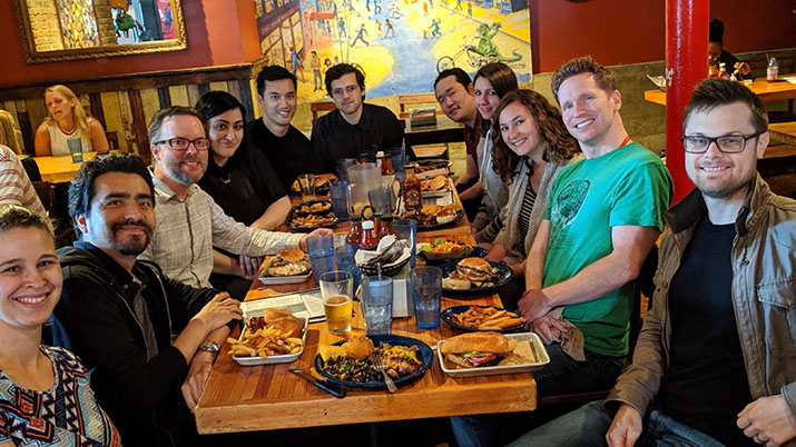
Fun Times at the Cole Lab!


People in the Lab


Michael W. Cole, PhD Principal Investigator

Associate Professor
Center for Molecular and Behavioral Neuroscience (CMBN), Rutgers University-Newark
Email: michael.cole@rutgers.edu

Michael's Rutgers University profile
Michael's curriculum vitae
Michael's citations on Google Scholar
Twitter: @TheColeLab


Ravi Mill, PhD Research Faculty

PhD: University of St. Andrews in Psychology & Neuroscience (2015)
BS: University College London in Psychology (2010)
Email: ravi.mill@rutgers.edu
Twitter: @RaviMill


Luke Hearne, PhD Postdoctoral Fellow

PhD: University of Queensland in Cognitive Neuroscience (2017)
BS: University of Queensland in Psychology (2012)
Luke's citations on Google Scholar
Email: luke.hearne@rutgers.edu
Twitter: @LukeJHearne


Ruben Sanchez-Romero, PhD Postdoctoral Fellow

PhD: Carnegie Mellon University (2018)
Ruben's citations on Google Scholar
Email: ruben.saro@rutgers.edu

Twitter: @rubensaro


Matthew Singh, PhD Postdoctoral Fellow

PhD: Washington University in St. Louis in Neuroscience (2020)
BA: University of Tennessee, Knoxville in Psychology, Mathematics & Neuroscience minor (2014)
Email: f.singh@wustl.edu


0

Kirsten Peterson Graduate Student

BS: University of Southern California in Computational Neuroscience (2018)
Email: klp173@rutgers.edu


0

Lakshman Chakravarthula Graduate Student

MS: Indian Institute of Technology Gandhinagar in Cognitive Science (2019)
Bachelor of Technology: Indian Institute of Technology Roorkee in Metallurgical and Materials Engineering (2017)


0

Alexandros Tzalavras Graduate Student

MS: New Jersey Institute of Technology in Biomedical Engineering (2022)
BS & MS: National Technical University of Athens (NTUA) – Athens, Greece in Electrical and Computer Engineering (2020)


0

Arun Aryal Graduate Student

BS: New Jersey Institute of Technology in Biomedical Engineering (2023)


Other members of the lab

Alisson López-Donado Undergraduate Research Assistant

Gifty Jones GS-LSAMP Undergraduate Research Assistant

Shraeyah N. Rajeshwaran Undergraduate Research Assistant

Cindy Hussein Undergraduate Research Assistant


Lab Alumni

Ella Podvalny Postdoctoral Fellow

Adeola Ajiboro GS-LSAMP Undergraduate Research Assistant

Temitope F. Ayetan Undergraduate Research Assistant

Carrisa Cocuzza Graduate Student

Micah Ketola Graduate Student

Edwin Lotero GS-LSAMP Undergraduate Research Assistant

Nicole Lalta GS-LSAMP Lab Manager & Research Assistant

Aditya Rao Research Assistant

Akshay Warrier Undergraduate Research Assistant

Avi Shah Research Assistant

Brian Keane Visiting Scholar

Jada White GS-LSAMP Undergraduate Research Assistant

Willio Jeanvilma GS-LSAMP Undergraduate Research Assistant

Richard Chen Graduate Student

Ethan McCormick Postdoctoral Fellow

Emily Winfield Lab Manager & Research Assistant

Takuya Ito Graduate Student

Diana Rodas GS-LSAMP Undergraduate Research Assistant

Catalina Guzman GS-LSAMP Undergraduate Research Assistant

Katelyn Arnemann Postdoctoral Fellow

Julia Hamilton Lab Manager & Research Assistant

Ian Kim Rotation Graduate Student

Marjolein Spronk Postdoctoral fellow

Aisha Assaf Undergraduate Research Assistant

Doug Schultz Postdoctoral fellow

Katherine Wolfert Rotation Graduate Student

Yanira Sanchez GS-LSAMP Undergraduate Research Assistant

Guochun Yang Visiting Scholar

Kaustubh Kulkarni Lab Manager & Research Assistant

Miguel Vivar Undergraduate Research Assistant

Brayan Zambrono Undergraduate Research Assistant

Levi Solomyak Lab Manager & Research Assistant

Senne Braem Visiting Scholar

Sara Horton Undergraduate Research Assistant

Eryn Fox Research Assistant

Pinelopi Kyriazi Rotation Graduate Student

Mia Tharp Visiting Scholar


Public Information & Outreach

On the Web:

In the news:


Publications

Preprints

Peterson KL, Sanchez-Romero R, Mill RD, Cole MW (Preprint). "Regularized partial correlation provides reliable functional connectivity estimates while correcting for widespread confounding". bioRxiv. doi:10.1101/2023.06.27.546751 On Twitter

Functional connectivity (FC) has been invaluable for understanding the brain's communication network, with strong potential for enhanced FC approaches to yield additional insights. Unlike with the fMRI field-standard method of pairwise correlation, theory suggests that partial correlation can estimate FC without confounded and indirect connections. However, partial correlation FC can also display low repeat reliability, impairing the accuracy of individual estimates. We hypothesized that reliability would be increased by adding regularization, which can reduce overfitting to noise in regression-based approaches like partial correlation. We therefore tested several regularized alternatives - graphical lasso, graphical ridge, and principal component regression - against unregularized partial and pairwise correlation, applying them to empirical resting-state fMRI and simulated data. As hypothesized, regularization vastly improved reliability, quantified using between-session similarity and intraclass correlation. This enhanced reliability then granted substantially more accurate individual FC estimates when validated against structural connectivity (empirical data) and ground truth networks (simulations). Graphical lasso showed especially high accuracy among regularized approaches, seemingly by maintaining more valid underlying network structures. We additionally found graphical lasso to be robust to noise levels, data quantity, and subject motion - common fMRI error sources. Lastly, we demonstrated that resting-state graphical lasso FC can effectively predict fMRI task activations and individual differences in behavior, further establishing its reliability, external validity, and ability to characterize task-related functionality. We recommend graphical lasso or similar regularized methods for calculating FC, as they can yield more valid estimates of unconfounded connectivity than field-standard pairwise correlation, while overcoming the poor reliability of unregularized partial correlation.

Mill RD, Cole MW (Preprint). "Neural representation dynamics reveal computational principles of cognitive task learning". bioRxiv. doi:10.1101/2023.06.27.546751 On Twitter

During cognitive task learning, neural representations must be rapidly constructed for novel task performance, then optimized for robust practiced task performance. How the geometry of neural representations changes to enable this transition from novel to practiced performance remains unknown. We hypothesized that practice involves a shift from compositional representations (task-general activity patterns that can be flexibly reused across tasks) to conjunctive representations (task-specific activity patterns specialized for the current task). Functional MRI during learning of multiple complex tasks substantiated this dynamic shift from compositional to conjunctive representations, which was associated with reduced cross-task interference (via pattern separation) and behavioral improvement. Further, we found that conjunctions originated in subcortex (hippocampus and cerebellum) and slowly spread to cortex, extending multiple memory systems theories to encompass task representation learning. The formation of conjunctive representations hence serves as a computational signature of learning, reflecting cortical-subcortical dynamics that optimize task representations in the human brain.

Cocuzza CV, Sanchez-Romero R, Ito T, Mill RD, Keane BP, Cole MW (Preprint). "Distributed network flows generate localized category selectivity in human visual cortex". bioRxiv. doi:10.1101/2022.02.19.481103 On Twitter

A central goal of neuroscience is to understand how function-relevant brain activations are generated. Here we test the hypothesis that function-relevant brain activations are generated primarily by distributed network flows. We focused on visual processing in human cortex, given the long-standing literature supporting the functional relevance of brain activations in visual cortex regions exhibiting visual category selectivity. We began by using fMRI data from N=352 human participants to identify category-specific responses in visual cortex for images of faces, places, body parts, and tools. We then systematically tested the hypothesis that distributed network flows can generate these localized visual category selective responses. This was accomplished using a recently developed approach for simulating — in a highly empirically constrained manner — the generation of task-evoked brain activations by modeling activity flowing over intrinsic brain connections. We next tested refinements to our hypothesis, focusing on how stimulus-driven network interactions initialized in V1 generate downstream visual category selectivity. We found evidence that network flows directly from V1 were sufficient for generating visual category selectivity, but that additional, globally distributed (whole-cortex) network flows increased category selectivity further. Using null network architectures we also found that each region's unique intrinsic "connectivity fingerprint" was key to the generation of category selectivity. These results generalized across regions associated with all four visual categories tested (bodies, faces, places, and tools), and provide evidence that the human brain's intrinsic network organization plays a prominent role in the generation of functionally relevant, localized responses.

Blujus J, Cole MW, Festa EK, Buka SL, Salloway SP, Heindel WC, Oh H (Preprint). Functional Redundancy of the Posterior Hippocampi, but not Anterior Hippocampi or Left Frontal Cortex, is Disrupted in Pathological Brain Aging. bioRxiv

As prevalence rates of Alzheimer’s disease (AD), the leading cause of dementia, are projected to more than double by 2050, emphasis has been placed on early intervention strategies that target resilience mechanisms to delay or prevent the onset of clinical symptoms. Several neural mechanisms underlying brain resilience to AD have been proposed, including redundant neural connections between the posterior hippocampi (HC) and all other brain regions, and global functional connectivity of the left frontal cortex (LFC). It remains unknown, however, if regional redundancy of the HC and LFC underscores neural resilience in the presence of AD pathologies. From the ADNI database, 363 cognitively normal older adults (CN) (N = 220; 36% Aβ+) and patients with Mild Cognitive Impairment (MCI) (N = 143; 51% Aβ+) were utilized. Regional redundancy was calculated from resting state fMRI data using a graph theoretical approach by summing the direct and indirect paths (path lengths=1-4) between each ROI and its 262 functional connections. The results showed that Aβ-status significantly disrupted posterior HC, but not anterior HC or LFC, redundancy. Aβ- groups showed higher redundancy of the bilateral posterior HC than Aβ+. In regard to redundancy-cognition relationships, higher posterior HC redundancy was related to better episodic memory performance, an effect which was primarily driven by the Aβ- group. Despite the positive relationship between posterior HC redundancy and cognition, we did not find compelling evidence that redundancy of the posterior HC serves in a resilience manner, as posterior HC redundancy did not moderate the potentially deleterious relationship between Aβ deposition and cognition. No relationships were found between anterior HC or LFC redundancy and cognitive performance. Together, these findings suggest that redundancy of the LFC does not underpin its role in resilience and that posterior HC redundancy may capture disruptions to network connectivity that occur as a result of Aβ deposition.

Podvalny E, Sanchez-Romero R, Cole MW (Preprint). Integrity of arousal-regulating brain circuitry predicts human cognitive abilities. bioRxiv doi.org/10.1101/2024.01.09.574917

Arousal state is regulated by the brainstem nucleus locus coeruleus, which sends wide-reaching projections to cortex. How this powerful neuromodulatory system is recruited to aid cognition is largely underexplored. Here, we hypothesized that select cortical regions may activate the arousal system, which in turn modulates large-scale brain activity, creating a circuit whose integrity predicts cognitive ability. We utilized the Human Connectome Project 7T functional magnetic resonance imaging dataset (N=149), acquired at rest with simultaneous eye tracking, along with extensive cognitive assessment for each subject. First, we discovered select frontoparietal cortical regions that likely drive large-scale spontaneous brain activity specifically via engaging the arousal system. Second, we show that the integrity of the arousal circuit driven by bilateral posterior cingulate cortex (associated with the default mode network) predicts subjects' cognitive abilities. This suggests that a cortical region that is typically associated with self-referential processing supports cognition by regulating the arousal system.

Chai M, Palenciano AF, Mill R, Cole MW, Braem S (Preprint). It is hard to prepare for task novelty: Cueing the novelty of upcoming tasks does not facilitate task performance. PsyArXiv 10.31234/osf.io/784du

Rapidly learning new tasks, such as using new technology or playing a new game,is ubiquitous in our daily lives. Previous studies suggest that our brain relies on different networks for rapid task learning versus retrieving known tasks from memory, and behavioralstudies have shown that novel versus practicedtasks benefit from different task preparation strategiesor control modes. Here, we investigatedwhether explicitly informingabout the novelty of anincoming task would help participants pre-adjust their task preparation strategies. We hypothesized that, if participants adopted differential preparatory strategies on novel versus practiced tasks, a task type cue informing about the novelty of the upcoming task couldhelp them adjust the correspondingcontrolmodein advance, leading to better task performance. Acrossfourexperiments, participants werefirsttrained on a subset of tasks, followed byatest session in whichthe task type cues were provided in some blocks but not others. After comparing task performance between cued anduncued blocks, our results indicated no benefitof cueingforbothpracticed and novel tasks, suggesting that peoplecannot pre-adjust their control mode in the absence of concrete task information.

Cole MW (Preprint). Cognitive flexibility as the shifting of brain network flows by flexible neural representations. PsyArXiv 10.31234/osf.io/kh4st

Our ability to overcome habitual responses in favor of goal-driven novel responses depends on frontoparietal cognitive control networks (CCNs). Recent and ongoing work is revealing the brain network and information processes that allow CCNs to generate cognitive flexibility. First, working memory processes necessary for flexible maintenance and manipulation of goal-relevant representations were recently found to depend on short-term network plasticity (in contrast to persistent activity) within CCN regions. Second, compositional (i.e., abstract and reusable) rule representations maintained within CCNs have been found to reroute network activity flows from stimulus to response, enabling flexible behavior. Together these findings suggest cognitive flexibility is enhanced by CCN-coordinated network mechanisms, utilizing compositional reuse of neural representations and network flows to flexibly accomplish task goals.

2024

Cole MW (In Press). "The explanatory power of activity flow models of brain function". Chapter in forthcoming book Computational and Network Modeling of Neuroimaging Data.

Tremendous neuroscientific progress has recently been made by mapping brain connectivity, complementing extensive knowledge of task-evoked brain activation patterns. However, despite evidence that they are related, these connectivity and activity lines of research have mostly progressed separately. Here I review the notable productivity and future promise of combining connectivity and task-evoked activity estimates into activity flow models. These data-driven computational models simulate the generation of task-evoked activations (including those linked to behavior), producing empirically-supported explanations of the origin of neurocognitive functions based on the flow of task-evoked activity over empirical brain connections. Critically, by incorporating causal principles and extensive empirical constraints from brain data, this approach can provide more mechanistic accounts of neurocognitive phenomena than purely predictive (as opposed to explanatory) models or models optimized primarily for task performance (e.g., standard artificial neural networks). The variety of activity-flow-based explanations reported so far are covered here along with important methodological and theoretical considerations when discovering new activity-flow-based explanations. Together, these considerations illustrate the promise of activity flow modeling for the future of neuroscience and ultimately for the development of novel clinical treatments (e.g., using brain stimulation) for brain disorders.

2023

Rosenberg-Lee M, Varma S, Cole MW, Abreu-Mendoza RA (2023). "Competing numerical magnitude codes in decimal comparison: Whole number and rational number distance both impact performance". Cognition. doi:10.1016/j.cognition.2023.105608

A critical difference between decimal and whole numbers is that among whole numbers the number of digits provides reliable information about the size of the number, e.g., double-digit numbers are larger than single-digit numbers. However, for decimals, fewer digits can sometimes denote a larger number (i.e., 0.8 > 0.27). Accordingly, children and adults perform worse when comparing such Inconsistent decimal pairs relative to Consistent pairs, where the larger number also has more digits (i.e., 0.87 > 0.2). Two explanations have been posited for this effect. The string length congruity account proposes that participants compare each position in the place value system, and they additionally compare the number of digits. The semantic interference account suggests that participants additionally activate the whole number referents of numbers – the numbers unadorned with decimal points (e.g., 8 < 27) – and compare these. The semantic interference account uniquely predicts that for Inconsistent problems with the same actual rational distance, those with larger whole number distances should be harder, e.g., 0.9 vs. 0.81 should be harder than 0.3 vs. 0.21 because 9 < < 81 whereas 3 < 21. Here we test this prediction in two experiments with college students (Study 1: n = 58 participants, Study 2: n = 78). Across both, we find a main effect of consistency, demonstrating string length effects, and also that whole number distance interferes with processing conflicting decimals, demonstrating semantic interference effects. Evidence for both effects supports the semantic interference account, highlighting that decimal comparison difficulties arise from multiple competing numerical codes. Finally, for accuracy we found no relationship between whole number distance sensitivity and math achievement, indicating that whole number magnitude interference affects participants similarly across the spectrum of math achievement.

Sanchez-Romero R, Ito T, Mill RD, Hanson SJ, Cole MW (2023). "Causally informed activity flow models provide mechanistic insight into network-generated cognitive activations". NeuroImage. 278:120300. doi:10.1016/j.neuroimage.2023.120300 Code On Twitter

Brain activity flow models estimate the movement of task-evoked activity over brain connections to help explain network-generated task functionality. Activity flow models have been shown to accurately generate task-evoked brain activations across a wide variety of brain regions and task conditions. However, these models have had limited explanatory power, given known issues with causal interpretations of the standard functional connectivity measures used to parameterize activity flow models. We show here that functional/effective connectivity (FC) measures grounded in causal principles facilitate mechanistic interpretation of activity flow models. We progress from simple to complex FC measures, with each adding algorithmic details reflecting causal principles. This reflects many neuroscientists’ preference for reduced FC measure complexity (to minimize assumptions, minimize compute time, and fully comprehend and easily communicate methodological details), which potentially trades off with causal validity. We start with Pearson correlation (the current field standard) to remain maximally relevant to the field, estimating causal validity across a range of FC measures using simulations and empirical fMRI data. Finally, we apply causal-FC-based activity flow modeling to a dorsolateral prefrontal cortex region (DLPFC), demonstrating distributed causal network mechanisms contributing to its strong activation during a working memory task. Notably, this fully distributed model is able to account for DLPFC working memory effects traditionally thought to rely primarily on within-region (i.e., not distributed) recurrent processes. Together, these results reveal the promise of parameterizing activity flow models using causal FC methods to identify network mechanisms underlying cognitive computations in the human brain.

Keane BP, Krekelberg B, Mill RD, Silverstein SM, Thompson JL, Serody MR, Barch DM, Cole MW (In Press). "Dorsal attention network activity during perceptual organization is distinct in schizophrenia and predictive of cognitive disorganization". European Journal of Neuroscience. doi:10.1111/ejn.15889 On Twitter

Visual shape completion is a canonical perceptual organization process that integrates spatially distributed edge information into unified representations of objects. People with schizophrenia show difficulty in discriminating completed shapes but the brain networks and functional connections underlying this perceptual difference remain poorly understood. Also unclear is whether brain network differences in schizophrenia occur in related illnesses or vary with illness features transdiagnostically. To address these topics, we scanned (fMRI) people with schizophrenia, bipolar disorder, or no psychiatric illness during rest and during a task in which they discriminated configurations that formed or failed to form completed shapes (illusory and fragmented condition, respectively). Multivariate pattern differences were identified on the cortical surface using 360 predefined parcels and 12 functional networks composed of such parcels. Brain activity flow mapping was used to evaluate the likely involvement of resting-state connections for shape completion. Illusory/fragmented task activation differences (“modulations”) in the dorsal attention network (DAN) could distinguish people with schizophrenia from the other groups (AUCs>.85) and could transdiagnostically predict cognitive disorganization severity. Activity flow over functional connections from the DAN could predict secondary visual network modulations in each group, except in schizophrenia. The secondary visual network was strongly and similarly modulated in each group. Task modulations were dispersed over more networks in patients compared to controls. In summary, DAN activity during visual perceptual organization is distinct in schizophrenia, symptomatically relevant, and potentially related to improper attention-related feedback into secondary visual areas.

2022

Hwang K, Shine JM, Cole MW, Sorenson E. (2022). "Thalamocortical contributions to cognitive task activity". eLife. doi:10.7554/eLife.81282

Thalamocortical interaction is a ubiquitous functional motif in the mammalian brain. Previously (Hwang et al., 2021), we reported that lesions to network hubs in the human thalamus are associated with multi-domain behavioral impairments in language, memory, and executive functions. Here, we show how task-evoked thalamic activity is organized to support these broad cognitive abilities. We analyzed functional magnetic resonance imaging (MRI) data from human subjects that performed 127 tasks encompassing a broad range of cognitive representations. We first investigated the spatial organization of task-evoked activity and found a basis set of activity patterns evoked to support processing needs of each task. Specifically, the anterior, medial, and posterior-medial thalamus exhibit hub-like activity profiles that are suggestive of broad functional participation. These thalamic task hubs overlapped with network hubs interlinking cortical systems. To further determine the cognitive relevance of thalamic activity and thalamocortical functional connectivity, we built a data-driven thalamocortical model to test whether thalamic activity can be used to predict cortical task activity. The thalamocortical model predicted task-specific cortical activity patterns, and outperformed comparison models built on cortical, hippocampal, and striatal regions. Simulated lesions to low-dimensional, multi-task thalamic hub regions impaired task activity prediction. This simulation result was further supported by profiles of neuropsychological impairments in human patients with focal thalamic lesions. In summary, our results suggest a general organizational principle of how the human thalamocortical system supports cognitive task activity.

Mill RD, Hamilton JL, Winfield EC, Lalta N, Chen RH, Cole MW (2022). "Network modeling of dynamic brain interactions predicts emergence of neural information that supports human cognitive behavior". PLOS Biology. doi:10.1101/2021.01.26.428276 Commentary on paper Code On Twitter

How cognitive task behavior is generated by brain network interactions is a central question in neuroscience. Answering this question calls for the development of novel analysis tools that can firstly capture neural signatures of task information with high spatial and temporal precision (the “where and when”) and then allow for empirical testing of alternative network models of brain function that link information to behavior (the “how”). We outline a novel network modeling approach suited to this purpose that is applied to noninvasive functional neuroimaging data in humans. We first dynamically decoded the spatiotemporal signatures of task information in the human brain by combining MRI-individualized source electroencephalography (EEG) with multivariate pattern analysis (MVPA). A newly developed network modeling approach—dynamic activity flow modeling—then simulated the flow of task-evoked activity over more causally interpretable (relative to standard functional connectivity [FC] approaches) resting-state functional connections (dynamic, lagged, direct, and directional). We demonstrate the utility of this modeling approach by applying it to elucidate network processes underlying sensory–motor information flow in the brain, revealing accurate predictions of empirical response information dynamics underlying behavior. Extending the model toward simulating network lesions suggested a role for the cognitive control networks (CCNs) as primary drivers of response information flow, transitioning from early dorsal attention network-dominated sensory-to-response transformation to later collaborative CCN engagement during response selection. These results demonstrate the utility of the dynamic activity flow modeling approach in identifying the generative network processes underlying neurocognitive phenomena.

Ito T, Klinger T, Schultz DH, Murray JD, Cole MW, Rigotti M (2022). "Compositional generalization through abstract representations in human and artificial neural networks". NeurIPS Proceedings. doi:10.48550/arXiv.2209.07431

Humans have a remarkable ability to rapidly generalize to new tasks that is difficult to reproduce in artificial learning systems. Compositionality has been proposed as a key mechanism supporting generalization in humans, but evidence of its neural implementation and impact on behavior is still scarce. Here we study the computational properties associated with compositional generalization in both humans and artificial neural networks (ANNs) on a highly compositional task. First, we identified behavioral signatures of compositional generalization in humans, along with their neural correlates using whole-cortex functional magnetic resonance imaging (fMRI) data. Next, we designed pretraining paradigms aided by a procedure we term "primitives pretraining" to endow compositional task elements into ANNs. We found that ANNs with this prior knowledge had greater correspondence with human behavior and neural compositional signatures. Importantly, primitives pretraining induced abstract internal representations, excellent zero-shot generalization, and sample-efficient learning. Moreover, it gave rise to a hierarchy of abstract representations that matched human fMRI data, where sensory rule abstractions emerged in early sensory areas, and motor rule abstractions emerged in later motor areas. Our findings give empirical support to the role of compositional generalization in human behavior, implicate abstract representations as its neural implementation, and illustrate that these representations can be embedded into ANNs by designing simple and efficient pretraining procedures.

Ito T, Yang GB, Laurent P, Schultz DH, Cole MW (2022). "Constructing neural network models from brain data reveals representational transformations linked to adaptive behavior". Nature Communications. 13, 673. doi:10.1038/s41467-022-28323-7 Code On Twitter

The human ability to adaptively implement a wide variety of tasks is thought to emerge from the dynamic transformation of cognitive information. We hypothesized that these transformations are implemented via conjunctive activations in “conjunction hubs”—brain regions that selectively integrate sensory, cognitive, and motor activations. We used recent advances in using functional connectivity to map the flow of activity between brain regions to construct a task-performing neural network model from fMRI data during a cognitive control task. We verified the importance of conjunction hubs in cognitive computations by simulating neural activity flow over this empirically-estimated functional connectivity model. These empirically-specified simulations produced above-chance task performance (motor responses) by integrating sensory and task rule activations in conjunction hubs. These findings reveal the role of conjunction hubs in supporting flexible cognitive computations, while demonstrating the feasibility of using empirically-estimated neural network models to gain insight into cognitive computations in the human brain.

Cocuzza CV, Sanchez-Romero R, Cole MW (2022). "Protocol for activity flow mapping of neurocognitive computations using the Brain Activity Flow Toolbox". STAR Protocols. 3, 1. doi:10.1016/j.xpro.2021.101094 Code On Twitter

Traditional cognitive neuroscience uses task-evoked activations to map neurocognitive processes (and information) to brain regions; however, how those processes are generated is unknown. We developed activity flow mapping to identify and empirically validate network mechanisms underlying the generation of neurocognitive processes. This approach models the movement of task-evoked activity over brain connections to predict task-evoked activations. We present a protocol for using the Brain Activity Flow Toolbox (https://colelab.github.io/ActflowToolbox/) to identify network mechanisms underlying neurocognitive processes of interest. For complete details on the use and execution of this protocol, please refer to Cole et al., 2021.

McCormick EM*, Arnemann KL*, Ito T, Hanson SJ, Cole MW (2022). "Latent functional connectivity underlying multiple brain states". Network Neuroscience; 6 (2): 570–590 doi:10.1162/netn_a_00234 [* = equal contribution] Code On Twitter

Functional connectivity (FC) studies have predominantly focused on resting state, where ongoing dynamics are thought to reflect the brain’s intrinsic network architecture; thought to be broadly relevant because it persists across brain states (i.e., state-general). However, it is unknown whether resting state is the optimal state for measuring intrinsic FC. We propose that latent FC, reflecting shared connectivity patterns across many brain states, better captures state-general intrinsic FC relative to measures derived from resting state alone. We estimated latent FC independently for each connection using leave-one-task-out factor analysis in 7 highly distinct task states (24 conditions) and resting state using fMRI data from the Human Connectome Project. Compared to resting-state connectivity, latent FC improves generalization to held-out brain states, better explaining patterns of connectivity and task-evoked activation. We also found that latent connectivity improved prediction of behavior outside the scanner, indexed by the general intelligence factor (g). Our results suggest that FC patterns shared across many brain states, rather than just resting state, better reflects state-general connectivity. This affirms the notion of “intrinsic” brain network architecture as a set of connectivity properties persistent across brain states, providing an updated conceptual and mathematical framework of intrinsic connectivity as a latent factor.

Schultz DH, Ito T, Cole MW (2022). "Global connectivity fingerprints predict the domain generality of multiple-demand regions". Cerebral Cortex, 32, 4464–4479 doi:10.1093/cercor/bhab495 On Twitter

A set of distributed cognitive control networks are known to contribute to diverse cognitive demands, yet it is unclear how these networks gain this domain-general capacity. We hypothesized that this capacity is largely due to the particular organization of the human brain’s intrinsic network architecture. Specifically, we tested the possibility that each brain region’s domain generality is reflected in its level of global (hub-like) intrinsic connectivity, as well as its particular global connectivity pattern (connectivity fingerprint). Consistent with prior work, we found that cognitive control networks exhibited domain generality, as they represented diverse task context information covering sensory, motor response, and logic rule domains. Supporting our hypothesis, we found that the level of global intrinsic connectivity (estimated with resting-state fMRI) was correlated with domain generality during tasks. Further, using a novel information fingerprint mapping approach, we found that each cognitive control region’s unique rule response profile (information fingerprint) could be predicted based on its unique intrinsic connectivity fingerprint and the information content in non-cognitive control regions. Together these results suggest that the human brain’s intrinsic network architecture supports its ability to represent diverse cognitive task information, largely via the location of multiple-demand regions within the brain’s global network organization.

Singh MF, Cole MW, Braver TS, Ching S (2022). "Developing control-theoretic objectives for large-scale brain dynamics and cognitive enhancement". Annual Reviews in Control. doi.org/10.1016/j.arcontrol.2022.05.001

The development of technologies for brain stimulation provides a means for scientists and clinicians to directly actuate the brain and nervous system. Brain stimulation has shown intriguing potential in terms of modifying particular symptom clusters in patients and behavioral characteristics of subjects. The stage is thus set for optimization of these techniques and the pursuit of more nuanced stimulation objectives, including the modification of complex cognitive functions such as memory and attention. Control theory and engineering will play a key role in the development of these methods, guiding computational and algorithmic strategies for stimulation. In particular, realizing this goal will require new development of frameworks that allow for controlling not only brain activity, but also latent dynamics that underlie neural computation and information processing. In the current opinion, we review recent progress in brain stimulation and outline challenges and potential research pathways associated with exogenous control of cognitive function.

Singh MF, Wang A, Cole MW, Ching S, Braver TS (2022). "Enhancing Task fMRI Preprocessing via Individualized Model-Based Filtering of Intrinsic Activity Dynamics". NeuroImage. doi:10.1016/j.neuroimage.2021.118836

Brain responses recorded during fMRI are thought to reflect both rapid, stimulus-evoked activity and the propagation of spontaneous activity through brain networks. In the current work, we describe a method to improve the estimation of task-evoked brain activity by first “filtering-out the intrinsic propagation of pre-event activity from the BOLD signal. We do so using Mesoscale Individualized NeuroDynamic (MINDy; Singh et al. 2020b) models built from individualized resting-state data to subtract the propagation of spontaneous activity from the task-fMRI signal (MINDy-based Filtering). After filtering, time-series are analyzed using conventional techniques. Results demonstrate that this simple operation significantly improves the statistical power and temporal precision of estimated group-level effects. Moreover, use of MINDy-based filtering increased the similarity of neural activation profiles and prediction accuracy of individual differences in behavior across tasks measuring the same construct (cognitive control). Thus, by subtracting the propagation of previous activity, we obtain better estimates of task-related neural effects.

2021

Hearne LJ, Mill RD, Keane BP, Repovs G, Anticevic A, Cole MW (2021). "Activity flow underlying abnormalities in brain activations and cognition in schizophrenia". Science Advances. 7(9) doi:10.1126/sciadv.abf2513 Code On Twitter

Cognitive dysfunction is a core feature of many brain disorders such as schizophrenia (SZ), and has been linked to both aberrant brain functional connectivity (FC) and aberrant cognitive brain activations. We propose that aberrant network activity flow over FC pathways leads to altered cognitive activations that produce cognitive dysfunction in SZ. We tested this hypothesis using activity flow mapping – an approach that models the movement of task-related activity between brain regions as a function of FC. Using fMRI data from SZ individuals and healthy controls during a working memory task, we found that activity flow models accurately predict aberrant cognitive activations across multiple brain networks. Within the same framework, we simulated a connectivity-based clinical intervention, predicting specific treatments that normalized brain activations and behavior in independent patients. Our results suggest that dysfunctional task-evoked activity flow is a large-scale network mechanism contributing to the emergence of cognitive dysfunction in SZ.

Mill RD, Winfield EC, Cole MW, Ray S (2021). "Structural MRI and functional connectivity features predict current clinical status and persistence behavior in prescription opioid users". NeuroImage: Clinical. 30. doi:10.1016/j.nicl.2021.102663

Prescription opioid use disorder (POUD) has reached epidemic proportions in the United States, raising an urgent need for diagnostic biological tools that can improve predictions of disease characteristics. The use of neuroimaging methods to develop such biomarkers have yielded promising results when applied to neurodegenerative and psychiatric disorders, yet have not been extended to prescription opioid addiction. With this long-term goal in mind, we conducted a preliminary study in this understudied clinical group. Univariate and multivariate approaches to distinguishing between POUD (n = 26) and healthy controls (n = 21) were investigated, on the basis of structural MRI (sMRI) and resting-state functional connectivity (restFC) features. Univariate approaches revealed reduced structural integrity in the subcortical extent of a previously reported addiction-related network in POUD subjects. No reliable univariate between-group differences in cortical structure or edgewise restFC were observed. Contrasting these mixed univariate results, multivariate machine learning classification approaches recovered more statistically reliable group differences, especially when sMRI and restFC features were combined in a multi-modal model (classification accuracy = 66.7%, p < .001). The same multivariate multi-modal approach also yielded reliable prediction of individual differences in a clinically relevant behavioral measure (persistence behavior; predicted-to-actual overlap r = 0.42, p = .009). Our findings suggest that sMRI and restFC measures can be used to reliably distinguish the neural effects of long-term opioid use, and that this endeavor numerically benefits from multivariate predictive approaches and multi-modal feature sets. This can serve as theoretical proof-of-concept for future longitudinal modeling of prognostic POUD characteristics from neuroimaging features, which would have clearer clinical utility.

Keane BP, Barch DM, Mill R, Silverstein SM, Krekelberg B, Cole MW (2021). "Brain network mechanisms of visual shape completion". NeuroImage. 236. doi:10.1101/2020.08.03.233403

Visual shape completion recovers object size, shape, and position from sparsely segregated edge elements. Studies of the process have largely focused on occipital cortex, but the role of other cortical areas and their functional interconnections remains poorly understood. To reveal the functional networks, connections, and regions of shape completion across the entire cortex, we scanned (fMRI) healthy adults during rest and during a task in which they indicated whether four pac-men formed a fat or thin illusory shape (illusory condition) or whether non-shape-forming pac-men were uniformly rotated left or right (fragmented condition). Task activation differences (illusory-fragmented), resting-state functional connectivity, and multivariate pattern analyses were performed on the cortical surface using 360 predefined cortical parcels (Glasser et al., 2016) and 12 functional networks composed of such parcels (Ji et al., 2019). Brain activity flow mapping (“ActFlow”) was used to evaluate the utility of resting-state connections for shape completion. Thirty-four parcels scattered across five functional networks were differentially active during shape completion. These regions were densely inter-connected during rest and a plurality occupied the secondary visual network. Posterior parietal, dorsolateral prefrontal, and orbitofrontal regions were also significant in the dorsal attention and frontoparietal networks. Functional connections from the dorsal attention network were key in modeling the emergence of activation differences (via ActFlow) in the secondary visual network and across all remaining networks. While shape completion is primarily driven by the secondary visual network, dorsal-attention regions are also involved, plausibly for relaying expectation-based signals about contour shape or position to ventral object-based areas.

Cole MW, Ito T, Cocuzza C, Sanchez-Romero R (2021). "The functional relevance of task-state functional connectivity". Journal of Neuroscience. 41(12):2684-2702. doi:10.1523/JNEUROSCI.1713-20.2021 Code On Twitter

Resting-state functional connectivity has provided substantial insight into intrinsic brain network organization, yet the functional importance of task-related change from that intrinsic network organization remains unclear. Indeed, such task-related changes are known to be small, suggesting they may have only minimal functional relevance. Alternatively, despite their small amplitude, these task-related changes may be essential for the human brain’s ability to adaptively alter its functionality via rapid changes in inter-regional relationships. We utilized activity flow mapping – an approach for building empirically-derived network models – to quantify the functional importance of task-state functional connectivity (above and beyond resting-state functional connectivity) in shaping cognitive task activations in the human brain. We found that task-state functional connectivity could be used to better predict independent fMRI activations across all 24 task conditions and all 360 cortical regions tested. Further, we found that prediction accuracy was strongly driven by individual-specific functional connectivity patterns, while functional connectivity patterns from other tasks (task-general functional connectivity) still improved predictions beyond resting-state functional connectivity. These findings suggest that task-related changes to functional connections play an important role in dynamically reshaping brain network organization, shifting the flow of neural activity during task performance.

Sanchez-Romero R, Cole MW (2021). "Combining multiple functional connectivity methods to improve causal inferences". Journal of Cognitive Neuroscience. 33(2):180–194. doi:10.1162/jocn_a_01580 On Twitter Code

Cognition and behavior emerge from brain network interactions, suggesting that causal interactions should be central to the study of brain function. Yet, approaches that characterize relationships among neural time series—functional connectivity (FC) methods—are dominated by methods that assess bivariate statistical associations rather than causal interactions. Such bivariate approaches result in substantial false positives because they do not account for confounders (common causes) among neural populations. A major reason for the dominance of methods such as bivariate Pearson correlation (with functional MRI) and coherence (with electrophysiological methods) may be their simplicity. Thus, we sought to identify an FC method that was both simple and improved causal inferences relative to the most popular methods. We started with partial correlation, showing with neural network simulations that this substantially improves causal inferences relative to bivariate correlation. However, the presence of colliders (common effects) in a network resulted in false positives with partial correlation, although this was not a problem for bivariate correlations. This led us to propose a new combined FC method (combinedFC) that incorporates simple bivariate and partial correlation FC measures to make more valid causal inferences than either alone. We release a toolbox for implementing this new combinedFC method to facilitate improvement of FC-based causal inferences. CombinedFC is a general method for FC and can be applied equally to resting-state and task-based paradigms.

Spronk M, Keane BP, Ito T, Kulkarni K, Ji JL, Anticevic A, Cole MW (2021). "A whole-brain and cross-diagnostic perspective on functional brain network dysfunction". Cerebral Cortex. 31(1):547-561. doi:10.1093/cercor/bhaa242 On Twitter

A wide variety of mental disorders have been associated with resting-state functional network alterations, which are thought to contribute to the cognitive changes underlying mental illness. These observations have seemed to support various theories postulating large-scale disruptions of brain systems in mental illness. However, existing approaches isolate differences in network organization without putting those differences in broad, whole-brain perspective. Using a graph distance measure - connectome-wide correlation - we found that whole-brain resting-state functional network organization in humans is highly similar across a variety of mental diseases and healthy controls. This similarity was observed across autism spectrum disorder, attention-deficit hyperactivity disorder, and schizophrenia. Nonetheless, subtle differences in network graph distance were predictive of diagnosis, suggesting that while functional connectomes differ little across health and disease those differences are informative. Such small network alterations may reflect the fact that most psychiatric patients maintain overall cognitive abilities similar to those of healthy individuals (relative to, e.g., the most severe schizophrenia cases), such that whole-brain functional network organization is expected to differ only subtly even for mental diseases with devastating effects on everyday life. These results suggest a need to reevaluate neurocognitive theories of mental illness, with a role for subtle functional brain network changes in the production of an array of mental diseases.

2020

Singh MF, Braver TS, Cole MW, Ching S (2020). "Estimation and validation of individualized dynamic brain models with resting state fMRI". NeuroImage. 221. doi:10.1016/j.neuroimage.2020.117046

A key challenge for neuroscience is to develop generative, causal models of the human nervous system in an individualized, data-driven manner. Previous initiatives have either constructed biologically-plausible models that are not constrained by individual-level human brain activity or used data-driven statistical characterizations of individuals that are not mechanistic. We aim to bridge this gap through the development of a new modeling approach termed Mesoscale Individualized Neurodynamic (MINDy) modeling, wherein we fit nonlinear dynamical systems models directly to human brain imaging data. The MINDy framework is able to produce these data-driven network models for hundreds to thousands of interacting brain regions in just 1–3 min per subject. We demonstrate that the models are valid, reliable, and robust. We show that MINDy models are predictive of individualized patterns of resting-state brain dynamical activity. Furthermore, MINDy is better able to uncover the mechanisms underlying individual differences in resting state activity than functional connectivity methods.

Ito T, Hearne LJ, Cole MW (2020). "A cortical hierarchy of localized and distributed processes revealed via dissociation of task activations, connectivity changes, and intrinsic timescales". NeuroImage. 221. doi:10.1016/j.neuroimage.2020.117141 On Twitter Code

Many studies have identified the role of localized and distributed cognitive functionality by mapping either local task-related activity or distributed functional connectivity (FC). However, few studies have directly explored the relationship between a brain region's localized task activity and its distributed task FC. Here we systematically evaluated the differential contributions of task-related activity and FC changes to identify a relationship between localized and distributed processes across the cortical hierarchy. We found that across multiple tasks, the magnitude of regional task-evoked activity was high in unimodal areas, but low in transmodal areas. In contrast, we found that task-state FC was significantly reduced in unimodal areas relative to transmodal areas. This revealed a strong negative relationship between localized task activity and distributed FC across cortical regions that was associated with the previously reported principal gradient of macroscale organization. Moreover, this dissociation corresponded to hierarchical cortical differences in the intrinsic timescale estimated from resting-state fMRI and region myelin content estimated from structural MRI. Together, our results contribute to a growing literature illustrating the differential contributions of a hierarchical cortical gradient representing localized and distributed cognitive processes.

Mill RD, Gordon BA, Balota DA, Cole MW (2020). "Predicting dysfunctional age-related task activations from resting-state network alterations". NeuroImage. 221. doi:10.1016/j.neuroimage.2020.117167 On Twitter Code

Alzheimer’s disease (AD) is linked to changes in fMRI task activations and fMRI resting-state functional connectivity (restFC), which can emerge early in the timecourse of illness. Study of these fMRI correlates of unhealthy aging has been conducted in largely separate subfields. Taking inspiration from neural network simulations, we propose a unifying mechanism wherein restFC network alterations associated with Alzheimer’s disease disrupt the ability for activations to flow between brain regions, leading to aberrant task activations. We apply this activity flow modeling framework in a large sample of clinically unimpaired older adults, which was segregated into healthy (low-risk) and at-risk subgroups based on established imaging (positron emission tomography amyloid) and genetic (apolipoprotein) risk factors for AD. We identified healthy task activations in individuals at low risk for AD, and then by estimating activity flow using at-risk AD restFC data we were able to predict the altered at-risk AD task activations. Thus, modeling the flow of healthy activations over at-risk AD connectivity effectively transformed the healthy aged activations into unhealthy aged activations. These results provide evidence that activity flow over altered intrinsic functional connections may act as a mechanism underlying Alzheimer’s-related dysfunction, even in very early stages of the illness. Beyond these mechanistic insights linking restFC with cognitive task activations, this approach has potential clinical utility as it enables prediction of task activations and associated cognitive dysfunction in individuals without requiring them to perform in-scanner cognitive tasks.

Ito T, Brincat SL, Siegel M, Mill RD, He BJ, Miller EK, Rotstein HG, and Cole MW (2020). "Task-Evoked Activity Quenches Neural Correlations and Variability in Large-Scale Brain Systems". PLOS Computational Biology. 16(8): e1007983. doi:10.1371/journal.pcbi.1007983 On Twitter

Many studies of large-scale neural systems have emphasized the importance of communication through increased inter-region correlations ("functional connectivity") during task states relative to resting state. In contrast, local circuit studies have demonstrated that task states reduce correlations among local neural populations, likely enhancing their information coding. Here we sought to adjudicate between these conflicting perspectives, assessing whether large-scale system correlations tend to increase or decrease during task states. To establish a mechanistic framework for interpreting changes in neural correlations, we conceptualized neural populations as having a sigmoidal neural transfer function. In a computational model we found that this straightforward assumption predicts reductions in neural populations' dynamic output range as task-evoked activity levels increase, reducing responsiveness to inputs from other regions (i.e., reduced correlations). We demonstrated this empirically in large-scale neural populations across two highly distinct data sets: human functional magnetic resonance imaging data and non-human primate spiking data. We found that task states increased global neural activity, while globally quenching neural variability and correlations. Further, this global reduction of neural correlations led to an overall increase in dimensionality (reflecting less information redundancy) during task states, providing an information-theoretic explanation for task-induced correlation reductions. Together, our results provide an integrative mechanistic account that encompasses measures of large-scale neural activity, variability, and correlations during resting and task states.

Cocuzza CV, Ito T, Schultz D, Bassett DS, Cole MW (2020). "Flexible coordinator and switcher hubs for adaptive task control". Journal of Neuroscience. 40(36):6949–6968. doi:10.1523/JNEUROSCI.2559-19.2020 On Twitter On Facebook Blog Post Code

Functional connectivity studies have identified at least two large-scale neural systems that constitute cognitive control networks – the frontoparietal network (FPN) and cingulo-opercular network (CON). Control networks are thought to support goal-directed cognition and behavior. It was previously shown that the FPN flexibly shifts its global connectivity pattern according to task goal, consistent with a “flexible hub” mechanism for cognitive control. Our aim was to build on this finding to develop a functional cartography (a multi-metric profile) of control networks in terms of dynamic network properties. We quantified network properties in (male and female) humans using a high-control-demand cognitive paradigm involving switching among 64 task sets. We hypothesized that cognitive control is enacted by the FPN and CON via distinct but complementary roles reflected in network dynamics. Consistent with a flexible “coordinator” mechanism, FPN connections were globally diverse across tasks, while maintaining within-network connectivity to aid cross-region coordination. Consistent with a flexible “switcher” mechanism, CON regions switched to other networks in a task-dependent manner, driven primarily by reduced within-network connections to other CON regions. This pattern of results suggests FPN acts as a dynamic, global coordinator of goal-relevant information, while CON transiently disbands to lend processing resources to other goal-relevant networks. This cartography of network dynamics reveals a dissociation between two prominent cognitive control networks, suggesting complementary mechanisms underlying goal-directed cognition.

Lamichhane B, Westbrook A, Cole MW, Braver T (2020). "Exploring brain-behavior relationships in the N-back task". NeuroImage. 212:1-11. doi:10.1016/j.neuroimage.2020.116683

Working memory (WM) function has traditionally been investigated in terms of two dimensions: within-individual effects of WM load, and between-individual differences in task performance. In human neuroimaging studies, the N-back task has frequently been used to study both. A reliable finding is that activation in frontoparietal regions exhibits an inverted-U pattern, such that activity tends to decrease at high load levels. Yet it is not known whether such U-shaped patterns are a key individual differences factor that can predict load-related changes in task performance. The current study investigated this question by manipulating load levels across a much wider range than explored previously (N = 1–6), and providing a more comprehensive examination of brain-behavior relationships. In a sample of healthy young adults (n = 57), the analysis focused on a distinct region of left lateral prefrontal cortex (LPFC) identified in prior work to show a unique relationship with task performance and WM function. In this region it was the linear slope of load-related activity, rather than the U-shaped pattern that was positively associated with individual differences in target accuracy. Comprehensive supplemental analyses revealed the brain-wide selectivity of this pattern. Target accuracy was also independently predicted by the global resting-state connectivity of this LPFC region. These effects were robust, as demonstrated by cross-validation analyses and out-of-sample prediction, and also critically, were primarily driven by the high-load conditions. Together, the results highlight the utility of high-load conditions for investigating individual differences in WM function.

Ito T, Hearne L, Mill R, Cocuzza C, Cole MW (2020). "Discovering the Computational Relevance of Brain Network Organization". Trends in Cognitive Sciences. 24, 25–38. doi:10.1016/j.tics.2019.10.005 On Twitter On Facebook

Understanding neurocognitive computations will require not just localizing cognitive information distributed throughout the brain but also determining how that information got there. We review recent advances in linking empirical and simulated brain network organization with cognitive information processing. Building on these advances, we offer a new framework for understanding the role of connectivity in cognition – network coding (encoding/decoding) models. These models utilize connectivity to specify the transfer of information via neural activity flow processes, successfully predicting the formation of cognitive representations in empirical neural data. The success of these models supports the possibility that localized neural functions mechanistically emerge (are computed) from distributed activity flow processes that are specified primarily by connectivity patterns.

Kar K, Ito T, Cole MW, and Krekelberg B (2020). "Transcranial Alternating Current Stimulation Attenuates BOLD Adaptation and Increases Functional Connectivity". Journal of Neurophysiology. 123: 428–438. doi:10.1152/jn.00376.2019 On Twitter

Transcranial alternating current stimulation (tACS) is used as a noninvasive tool for cognitive enhancement and clinical applications. The physiological effects of tACS, however, are complex and poorly understood. Most studies of tACS focus on its ability to entrain brain oscillations, but our behavioral results in humans and extracellular recordings in nonhuman primates support the view that tACS at 10 Hz also affects brain function by reducing sensory adaptation. Our primary goal in the present study is to test this hypothesis using blood oxygen level-dependent (BOLD) imaging in human subjects. Using concurrent functional magnetic resonance imaging (fMRI) and tACS, and a motion adaptation paradigm developed to quantify BOLD adaptation, we show that tACS significantly attenuates adaptation in the human motion area (hMT+). In addition, an exploratory analysis shows that tACS increases functional connectivity of the stimulated hMT+ with the rest of the brain and the dorsal attention network in particular. Based on field estimates from individualized head models, we relate these changes to the strength of tACS-induced electric fields. Specifically, we report that functional connectivity (between hMT+ and any other region of interest) increases in proportion to the field strength in the region of interest. These findings add support for the claim that weak 10-Hz currents applied to the scalp modulate both local and global measures of brain activity.

2019

Reid A, Headley DB, Mill RD, Sanchez-Romero R, Uddin LQ, Marinazzo D, Lurie DJ, Valdés-Sosa PA, Hanson SJ, Biswal BB, Calhoun V, Poldrack RA, Cole MW (2019). "Advancing functional connectivity research from association to causation". Nature Neuroscience. doi: 10.1038/s41593-019-0510-4 On Twitter On Facebook

Cognition and behavior emerge from brain network interactions, such that investigating causal interactions should be central to the study of brain function. Approaches that characterize statistical associations among neural time series—functional connectivity (FC) methods—are likely a good starting point for estimating brain network interactions. Yet only a subset of FC methods ('effective connectivity') is explicitly designed to infer causal interactions from statistical associations. Here we incorporate best practices from diverse areas of FC research to illustrate how FC methods can be refined to improve inferences about neural mechanisms, with properties of causal neural interactions as a common ontology to facilitate cumulative progress across FC approaches. We further demonstrate how the most common FC measures (correlation and coherence) reduce the set of likely causal models, facilitating causal inferences despite major limitations. Alternative FC measures are suggested to immediately start improving causal inferences beyond these common FC measures.

Yang GR, Cole MW, Rajan K (2019). "How to study the neural mechanisms of multiple tasks". Current Opinion in Behavioral Sciences. 29:134–143 doi.org/10.1016/j.cobeha.2019.07.001

Most biological and artificial neural systems are capable of completing multiple tasks. However, the neural mechanism by which multiple tasks are accomplished within the same system is largely unclear. We start by discussing how different tasks can be related, and methods to generate large sets of inter-related tasks to study how neural networks and animals perform multiple tasks. We then argue that there are mechanisms that emphasize either specialization or flexibility. We will review two such neural mechanisms underlying multiple tasks at the neuronal level (modularity and mixed selectivity), and discuss how different mechanisms can emerge depending on training methods in neural networks.

Bolt T, Nomi JS, Bainter S, Cole MW, Uddin LQ (2019). “The Situation or the Person? Individual and Task-Evoked Differences in BOLD Activity”. Human Brain Mapping. doi:10.1002/hbm.24570

Investigations of between‐person variability are enjoying a recent resurgence in functional magnetic resonance imaging (fMRI) research. Several recent studies have found persistent between‐person differences in blood‐oxygenated‐level dependent (BOLD) activation patterns and resting‐state functional connectivity. Conflicting findings have been reported regarding the extent to which (a) between‐person or (b) within‐person cognitive state differences explain differences in BOLD activation patterns. These discrepancies may arise due to statistical analysis choices, parcellation resolution, and limited sampling of task‐fMRI datasets. We attempt to address these issues in a large‐scale analysis of several task‐fMRI paradigms. Using a novel application of multivariate distance matrix regression, we examine between‐person and task‐condition variability estimates across varying levels of "resolution", from a coarse region‐of‐interest level to the vertex‐level, and across different distance metrics. These analyses revealed that under most circumstances, differences in task conditions explained a greater amount of variance in activation map differences than between‐person differences. However, this finding was reversed when comparing activation maps at a "high‐resolution" vertex level. More generally, we observed that when moving from "low" to "high" resolutions, the variance explained by between‐person differences increased while variance explained by task conditions decreased. We further analyzed the relationships among subject‐level activation maps across all task‐conditions using an unsupervised clustering approach and identified a superordinate task structure. This structure went beyond conventional task labels and highlighted those experimental manipulations across task conditions that produce contrasting versus similar whole‐brain activation patterns. Overall, these analyses suggest that the question of the subject‐ versus task‐effects on BOLD activation patterns is nontrivial, and depends on the comparison "resolution," choice of distance metric, and the coding of task‐conditions.

Cole MW, Ito T, Schultz D, Mill R, Chen R, Cocuzza C (2019). "Task activations produce spurious but systematic inflation of task functional connectivity estimates". NeuroImage. doi:10.1016/j.neuroimage.2018.12.054 On Twitter Code

Most neuroscientific studies have focused on task-evoked activations (activity amplitudes at specific brain locations), providing limited insight into the functional relationships between separate brain locations. Task-state functional connectivity (FC) – statistical association between brain activity time series during task performance – moves beyond task-evoked activations by quantifying functional interactions during tasks. However, many task-state FC studies do not remove the first-order effect of task-evoked activations prior to estimating task-state FC. It has been argued that this results in the ambiguous inference "likely active or interacting during the task", rather than the intended inference "likely interacting during the task". Utilizing a neural mass computational model, we verified that task-evoked activations substantially and inappropriately inflate task-state FC estimates, especially in functional MRI (fMRI) data. Various methods attempting to address this problem have been developed, yet the efficacies of these approaches have not been systematically assessed. We found that most standard approaches for fitting and removing mean task-evoked activations were unable to correct these inflated correlations. In contrast, methods that flexibly fit mean task-evoked response shapes effectively corrected the inflated correlations without reducing effects of interest. Results with empirical fMRI data confirmed the model's predictions, revealing activation-induced task-state FC inflation for both Pearson correlation and psychophysiological interaction (PPI) approaches. These results demonstrate that removal of mean task-evoked activations using an approach that flexibly models task-evoked response shape is an important preprocessing step for valid estimation of task-state FC.

Ji JL*, Spronk M*, Kulkarni K, Repovs G, Anticevic A**, Cole MW** (2019). "Mapping the human brain's cortical-subcortical functional network organization". NeuroImage. 185:35–57. doi:10.1016/j.neuroimage.2018.10.006 [* = equal contribution; ** = senior authors] On Facebook Brain Network Partition

Understanding complex systems such as the human brain requires characterization of the system's architecture across multiple levels of organization – from neurons, to local circuits, to brain regions, and ultimately large-scale brain networks. Here we focus on characterizing the human brain's large-scale network organization, as it provides an overall framework for the organization of all other levels. We developed a highly principled approach to identify cortical network communities at the level of functional systems, calibrating our community detection algorithm using extremely well-established sensory and motor systems as guides. Building on previous network partitions, we replicated and expanded upon well-known and recently-identified networks, including several higher-order cognitive networks such as a left-lateralized language network. We expanded these cortical networks to subcortex, revealing 358 highly-organized subcortical parcels that take part in forming whole-brain functional networks. Notably, the identified subcortical parcels are similar in number to a recent estimate of the number of cortical parcels (360). This whole-brain network atlas – released as an open resource for the neuroscience community – places all brain structures across both cortex and subcortex into a single large-scale functional framework, with the potential to facilitate a variety of studies investigating large-scale functional networks in health and disease.

Schultz DH, Ito T, Solomyak L, Chen RH, Mill RD, Anticevic A, Cole MW (2019). "Global connectivity of the frontoparietal cognitive control network is related to depression symptoms in the general population". Network Neuroscience. 3:1:107-123. doi:10.1162/NETN_a_00056 On Twitter On Facebook Blog Post

We all vary in our mental health, even among people not meeting diagnostic criteria for mental illness. Understanding this individual variability may reveal factors driving the risk for mental illness, as well as factors driving sub-clinical problems that still adversely affect quality of life. To better understand the large-scale brain network mechanisms underlying this variability we examined the relationship between mental health symptoms and resting-state functional connectivity patterns in cognitive control systems. One such system is the frontoparietal cognitive control network (FPN). Changes in FPN connectivity may impact mental health by disrupting the ability to regulate symptoms in a goal-directed manner. Here we test the hypothesis that FPN dysconnectivity relates to mental health symptoms even among individuals who do not meet formal diagnostic criteria but may exhibit meaningful symptom variation. We found that depression symptoms severity negatively correlated with between-network global connectivity (BGC) of the FPN. This suggests that decreased connectivity between the FPN and the rest of the brain is related to increased depression symptoms in the general population. These findings complement previous clinical studies to support the hypothesis that global FPN connectivity contributes to the regulation of mental health symptoms across both health and disease.

2018

Chen RH, Ito T, Kulkarni KR, Cole MW (2018). "The human brain traverses a common activation-pattern state space across task and rest". Brain Connectivity. 8:7. doi:10.1089/brain.2018.0586 On Twitter On Facebook

Much of our lives are spent in unconstrained rest states, yet cognitive brain processes are primarily investigated using task-constrained states. It may be possible to utilize the insights gained from experimental control of task processes as reference points for investigating unconstrained rest. To facilitate comparison of rest and task functional MRI data we focused on activation amplitude patterns, commonly used for task but not rest analyses. During rest, we identified spontaneous changes in temporally extended whole-brain activation-pattern states. This revealed a hierarchical organization of rest states. The top consisted of two competing states consistent with previously identified “task-positive” and “task-negative” activation patterns. These states were composed of more specific states that repeated over time and across individuals. Contrasting with the view that rest consists of only task-negative states, task-positive states occurred 40% of the time while individuals "rested", suggesting task-focused activity may occur during rest. Together our results suggest brain activation dynamics form a general hierarchy across task and rest, with a small number of dominant general states reflecting basic functional modes and a variety of specific states potentially reflecting a wide variety of cognitive processes.

Dixon ML, De La Vega A, Mills C, Andrews-Hanna J, Spreng RN, Cole MW, Christoff K (2018). "Heterogeneity Within the Frontoparietal Control Network and its Relationship to the Default and Dorsal Attention Networks". Proceedings of the National Academy of Sciences.

The frontoparietal control network (FPCN) plays a central role in executive control. It has been predominantly viewed as a unitary domain general system. Here, we examined patterns of FPCN functional connectivity (FC) across multiple conditions of varying cognitive demands, in order to test for FPCN heterogeneity. We identified two distinct subsystems within the FPCN based on hierarchical clustering and machine learning classification analyses of within-FPCN FC patterns. These two FPCN subsystems exhibited distinct patterns of FC the default network (DN) and the dorsal attentional network (DAN). This 2-fold FPCN differentiation was observed across four independent data sets, across 9 different conditions (rest and 8 tasks), as well as in meta-analytic co-activation patterns. The extent of FPCN differentiation varied across conditions, suggesting flexible adaptation to task demands. These findings reveal a flexible and heterogeneous FPCN organization that may in part emerge from separable DN and DAN processing streams.

Cole MW, Patrick L, Meiran N, Braver T (2018). "A role for proactive control in rapid instructed task learning". Acta Psychologica. 184:20–30. doi:10.1016/j.actpsy.2017.06.004 On Twitter

Humans are often remarkably fast at learning novel tasks from instructions. Such rapid instructed task learning (RITL) likely depends upon the formation of new associations between long-term memory representations, which must then be actively maintained to enable successful task implementation. Consequently, we hypothesized that RITL relies more heavily on a proactive mode of cognitive control, in which goal-relevant information is actively maintained in preparation for anticipated high control demands. We tested this hypothesis using a recently developed cognitive paradigm consisting of 60 novel tasks involving RITL and 4 practiced tasks, with identical task rules and stimuli used across both task types. A robust behavioral cost was found in novel relative to practiced task performance, which was present even when the two were randomly inter-mixed, such that task-switching effects were equated. Novelty costs were most prominent under time-limited preparation conditions. In self-paced conditions, increased preparation time was found for novel trials, and was selectively associated with enhanced performance, suggesting greater proactive control for novel tasks. These results suggest a key role for proactive cognitive control in the ability to rapidly learn novel tasks from instructions.

2017

Ito T, Kulkarni KR, Schultz DH, Mill RD, Chen RH, Solomyak LI, Cole MW (2017). "Cognitive task information is transferred between brain regions via resting-state network topology". Nature Communications. 8:1027. doi:10.1038/s41467-017-01000-w On Facebook Blog Post Code

Resting-state network connectivity has been associated with a variety of cognitive abilities, yet it remains unclear how these connectivity properties might contribute to the neurocognitive computations underlying these abilities. We developed a new approach—information transfer mapping—to test the hypothesis that resting-state functional network topology describes the computational mappings between brain regions that carry cognitive task information. Here, we report that the transfer of diverse, task-rule information in distributed brain regions can be predicted based on estimated activity flow through resting-state network connections. Further, we find that these task-rule information transfers are coordinated by global hub regions within cognitive control networks. Activity flow over resting-state connections thus provides a large-scale network mechanism for cognitive task information transfer and global information coordination in the human brain, demonstrating the cognitive relevance of resting-state network topology.

Cole, MW (2017). "Control and Connectivity: Dynamic Networks in the Human Brain". In The Wiley Handbook of Cognitive Control, edited by Tobias Egner, 314–33. Chichester, UK: John Wiley & Sons, Ltd. doi:10.1002/9781118920497.ch18.

The human brain is an extremely complex network consisting of billions of nodes and trillions of connections (Azevedo et al., 2009). Somehow, the complex spatiotemporal dynamics that play out on this network architecture produce cognitive control — a broad domain composed of goal‐directed thoughts and behaviours. Until recently, however, even those who conceptualised cognitive control in terms of brain network mechanisms nonetheless continued to exclusively use localisation tools such as single‐unit electrophysiology and functional MRI (fMRI) general linear models to investigate its neural basis. Here I will cover recent developments that are allowing for proper characterisation of the brain network basis of cognitive control. This includes methodological advances in characterising human brain connectivity, such as resting‐state and task‐state functional connectivity MRI and diffusion weighted imaging (DWI), as well as advances in identifying more descriptive network components such as brain hubs. Finally, I will cover theoretical insights gained from these advances, such as the nested organisation of cognitive control brain networks and the role of hub dynamics in implementing cognitive control functionality.

Mill R, Ito T, Cole M (2017). "From connectome to cognition: The search for mechanism in human functional brain networks". NeuroImage. doi:10.1016/j.neuroimage.2017.01.060 On Twitter On Facebook

Recent developments in functional connectivity research have expanded the scope of human neuroimaging, from identifying changes in regional activation amplitudes to detailed mapping of large-scale brain networks. However, linking network processes to a clear role in cognition demands advances in the theoretical frameworks, algorithms, and experimental approaches applied. This would help evolve the field from a descriptive to an explanatory state, by targeting network interactions that can mechanistically account for cognitive effects. In the present review, we provide an explicit framework to aid this search for “network mechanisms”, which anchors recent methodological advances in functional connectivity estimation to a renewed emphasis on careful experimental design. We emphasize how this framework can address specific questions in network neuroscience. These span ambiguity over the cognitive relevance of resting-state networks, how to characterize task-evoked and spontaneous network dynamics, how to identify directed or “effective” connections, and how to apply multivariate pattern analysis at the network level. In parallel, we apply the framework to highlight the mechanistic interaction of network components that remain “stable” across task domains and more “flexible” components associated with on-task reconfiguration. By emphasizing the need to structure the use of diverse analytic approaches with sound experimentation, our framework promotes an explanatory mapping between the workings of the cognitive mind and the large-scale network mechanisms of the human brain.

Cole MW, Braver T, Meiran N (2017). "The task novelty paradox: Flexible control of inflexible neural pathways during rapid instructed task learning". Neuroscience & Biobehavioral Reviews. doi:10.1016/j.neubiorev.2017.02.009 On Twitter On Facebook

Rapid instructed task learning (RITL) is one of the most remarkable human abilities, when considered from both computational and evolutionary perspectives. A key feature of RITL is that it enables new goals to be immediately pursued (and shared) following formation of task representations. Although RITL is a form of cognitive control that engenders immense flexibility, it also seems to produce inflexible activation of action plans in inappropriate contexts. We argue that this “prepared reflex” effect arises because RITL is implemented in the brain as a “flexible hub” mechanism, in which top-down influences from the frontoparietal control network reroute pathways among procedure-implementing brain areas (e.g., perceptual and motor areas). Specifically, we suggest that RITL-based proactive control – the preparatory biasing of task-relevant functional network routes – results in inflexible associative processing, demanding compensation in the form of increased reactive (in-the-moment) control. Thus, RITL produces a computational trade-off, in which the top-down influences of flexible hubs increase overall cognitive flexibility, but at the cost of temporally localized inflexibility (the prepared reflex effect).

Mill R, Bagic A, Bostan A, Schneider W, Cole MW (2017). "Empirical validation of directed functional connectivity". NeuroImage. 146:275–287. doi:10.1016/j.neuroimage.2016.11.037

Mapping directions of influence in the human brain connectome represents the next phase in understanding its functional architecture. However, a host of methodological uncertainties have impeded the application of directed connectivity methods, which have primarily been validated via 'ground truth' connectivity patterns embedded in simulated functional MRI (fMRI) and magneto-/electro-encephalography (MEG/EEG) datasets. Such simulations rely on many generative assumptions, and we hence utilized a different strategy involving empirical data in which a ground truth directed connectivity pattern could be anticipated with confidence. Specifically, we exploited the established 'sensory reactivation' effect in episodic memory, in which retrieval of sensory information reactivates regions involved in perceiving that sensory modality. Subjects performed a paired associate task in separate fMRI and MEG sessions, in which a ground truth reversal in directed connectivity between auditory and visual sensory regions was instantiated across task conditions. This directed connectivity reversal was successfully recovered across different algorithms, including Granger causality and Bayes network (IMAGES) approaches, and across fMRI ('raw' and deconvolved) and source-modeled MEG. These results extend simulation studies of directed connectivity, and offer practical guidelines for the use of such methods in clarifying causal mechanisms of neural processing.

2016

Cole MW, Ito T, Bassett DS, Schultz DH (2016). "Activity flow over resting-state networks shapes cognitive task activations". Nature Neuroscience. doi:10.1038/nn.4406 On Facebook Blog Post Code

Resting-state functional connectivity (FC) has helped reveal the intrinsic network organization of the human brain, yet its relevance to cognitive task activations has been unclear. Uncertainty remains despite evidence that resting-state FC patterns are highly similar to cognitive task activation patterns. Identifying the distributed processes that shape localized cognitive task activations may help reveal why resting-state FC is so strongly related to cognitive task activations. We found that estimating task-evoked activity flow (the spread of activation amplitudes) over resting-state FC networks allowed prediction of cognitive task activations in a large-scale neural network model. Applying this insight to empirical functional MRI data, we found that cognitive task activations can be predicted in held-out brain regions (and held-out individuals) via estimated activity flow over resting-state FC networks. This suggests that task-evoked activity flow over intrinsic networks is a large-scale mechanism explaining the relevance of resting-state FC to cognitive task activations.

Schultz DH & Cole MW (2016). "Higher Intelligence Is Associated with Less Task-Related Brain Network Reconfiguration". Journal of Neuroscience. doi:10.1523/JNEUROSCI.0358-16.2016

The human brain is able to exceed modern computers on multiple computational demands (e.g., language, planning) using a small fraction of the energy. The mystery of how the brain can be so efficient is compounded by recent evidence that all brain regions are constantly active as they interact in so-called resting-state networks (RSNs). To investigate the brain's ability to process complex cognitive demands efficiently, we compared functional connectivity (FC) during rest and multiple highly distinct tasks. We found previously that RSNs are present during a wide variety of tasks and that tasks only minimally modify FC patterns throughout the brain. Here, we tested the hypothesis that, although subtle, these task-evoked FC updates from rest nonetheless contribute strongly to behavioral performance. One might expect that larger changes in FC reflect optimization of networks for the task at hand, improving behavioral performance. Alternatively, smaller changes in FC could reflect optimization for efficient (i.e., small) network updates, reducing processing demands to improve behavioral performance. We found across three task domains that high-performing individuals exhibited more efficient brain connectivity updates in the form of smaller changes in functional network architecture between rest and task. These smaller changes suggest that individuals with an optimized intrinsic network configuration for domain-general task performance experience more efficient network updates generally. Confirming this, network update efficiency correlated with general intelligence. The brain's reconfiguration efficiency therefore appears to be a key feature contributing to both its network dynamics and general cognitive ability.

Schultz DH, Cole MW (2016) "Integrated Brain Network Architecture Supports Cognitive Task Performance". Neuron. 92:278–279. doi:10.1016/j.neuron.2016.10.004

Spontaneous fluctuations in neural activity and connectivity are thought to support cognition and behavior. In this issue of Neuron, Shine et al. (2016) describe a possible mechanism responsible for fluctuations in the human brain’s network architecture that are related to rapid shifts in cognitive state.

Cole M.W., Yang G. J., Murray J. D., Repovs G., Anticevic A. (2016). "Functional connectivity change as shared signal dynamics". Journal of Neuroscience Methods. doi:10.1016/j.jneumeth.2015.11.011

Background An increasing number of neuroscientific studies gain insights by focusing on differences in functional connectivity – between groups, individuals, temporal windows, or task conditions. We found using simulations that additional insights into such differences can be gained by forgoing variance normalization, a procedure used by most functional connectivity measures. Simulations indicated that these functional connectivity measures are sensitive to increases in independent fluctuations (unshared signal) in time series, consistently reducing functional connectivity estimates (e.g., correlations) even though such changes are unrelated to corresponding fluctuations (shared signal) between those time series. This is inconsistent with the common notion of functional connectivity as the amount of inter-region interaction.
New Method Simulations revealed that a version of correlation without variance normalization – covariance – was able to isolate differences in shared signal, increasing interpretability of observed functional connectivity change. Simulations also revealed cases problematic for non-normalized methods, leading to a “covariance conjunction” method combining the benefits of both normalized and non-normalized approaches.
Results We found that covariance and covariance conjunction methods can detect functional connectivity changes across a variety of tasks and rest in both clinical and non-clinical functional MRI datasets.
Comparison with Existing Method(s) We verified using a variety of tasks and rest in both clinical and non-clinical functional MRI datasets that it matters in practice whether correlation, covariance, or covariance conjunction methods are used.
Conclusions These results demonstrate the practical and theoretical utility of isolating changes in shared signal, improving the ability to interpret observed functional connectivity change.

Etzel J.A., Cole M.W., Zacks J.M., Kay K.N., Braver T.S. (2016). "Reward motivation enhances task coding in frontoparietal cortex". Cerebral Cortex. 26:4:1647-1659. doi.org/10.1093/cercor/bhu327

Reward motivation often enhances task performance, but the neural mechanisms underlying such cognitive enhancement remain unclear. Here, we used a multivariate pattern analysis (MVPA) approach to test the hypothesis that motivation-related enhancement of cognitive control results from improved encoding and representation of task set information. Participants underwent two fMRI sessions of cued task switching, the first under baseline conditions, and the second with randomly intermixed reward incentive and no-incentive trials. Information about the upcoming task could be successfully decoded from cue-related activation patterns in a set of frontoparietal regions typically associated with task control. More critically, MVPA classifiers trained on the baseline session had significantly higher decoding accuracy on incentive than non-incentive trials, with decoding improvement mediating reward-related enhancement of behavioral performance. These results strongly support the hypothesis that reward motivation enhances cognitive control, by improving the discriminability of task-relevant information coded and maintained in frontoparietal brain regions.

Cole M.W., Ito T., Braver T.S. (2016). "The behavioral relevance of task information in human prefrontal cortex". Cerebral Cortex. doi: 10.1093/cercor/bhv072

Human lateral prefrontal cortex (LPFC) is thought to play a critical role in enabling cognitive flexibility, particularly when performing novel tasks. However, it remains to be established whether LPFC representation of task-relevant information in such situations actually contributes to successful performance. We utilized pattern classification analyses of functional MRI activity to identify novelty-sensitive brain regions as participants rapidly switched between performance of 64 complex tasks, 60 of which were novel. In three of these novelty-sensitive regions – located within distinct areas of left anterior LPFC – trial-evoked activity patterns discriminated correct from error trials. Further, these regions also contained information regarding the task-relevant decision rule, but only for successfully performed trials. This suggests that left anterior LPFC may be particularly important for representing task information that contributes to the cognitive flexibility needed to perform successfully in novel task situations.

2015

Mattar M., Cole M.W., Thompson-Schill S., Bassett D.S. (2015). "A Functional Cartography of Cognitive Systems". PLOS Computational Biology. 11:12:e1004533. doi:10.1371/journal.pcbi.1004533.

One of the most remarkable features of the human brain is its ability to adapt rapidly and efficiently to external task demands. Novel and non-routine tasks, for example, are implemented faster than structural connections can be formed. The neural underpinnings of these dynamics are far from understood. Here we develop and apply novel methods in network science to quantify how patterns of functional connectivity between brain regions reconfigure as human subjects perform 64 different tasks. By applying dynamic community detection algorithms, we identify groups of brain regions that form putative functional communities, and we uncover changes in these groups across the 64-task battery. We summarize these reconfiguration patterns by quantifying the probability that two brain regions engage in the same network community (or putative functional module) across tasks. These tools enable us to demonstrate that classically defined cognitive systems—including visual, sensorimotor, auditory, default mode, fronto-parietal, cingulo-opercular and salience systems—engage dynamically in cohesive network communities across tasks. We define the network role that a cognitive system plays in these dynamics along the following two dimensions: (i) stability vs. flexibility and (ii) connected vs. isolated. The role of each system is therefore summarized by how stably that system is recruited over the 64 tasks, and how consistently that system interacts with other systems. Using this cartography, classically defined cognitive systems can be categorized as ephemeral integrators, stable loners, and anything in between. Our results provide a new conceptual framework for understanding the dynamic integration and recruitment of cognitive systems in enabling behavioral adaptability across both task and rest conditions. This work has important implications for understanding cognitive network reconfiguration during different task sets and its relationship to cognitive effort, individual variation in cognitive performance, and fatigue.

Cole M.W., Ito T., Braver T.S. (2015). "Lateral prefrontal cortex contributes to fluid intelligence via multi-network connectivity". Brain Connectivity. doi: 10.1089/brain.2015.0357

Our ability to effectively adapt to novel circumstances – as measured by general fluid intelligence – has recently been tied to the global connectivity of lateral prefrontal cortex (LPFC). Global connectivity is a broad measure that summarizes both within-network and across-network connectivity. We used additional graph theoretical measures to better characterize the nature of LPFC connectivity and its relationship with fluid intelligence. We specifically hypothesized that LPFC is a “connector” hub with across-network connectivity that contributes to fluid intelligence independently of within-network connectivity. We verified that LPFC was in the top 10% of brain regions in terms of across-network connectivity, suggesting it is a strong connector hub. Importantly, we found that LPFC across-network connectivity predicted individuals’ fluid intelligence, and that this correlation remained statistically significant when controlling for global connectivity (which includes within-network connectivity). This supports the conclusion that across-network connectivity independently contributes to the relationship between LPFC connectivity and intelligence. These results suggest LPFC contributes to fluid intelligence by being a connector hub with truly global multi-system connectivity throughout the brain.

Anticevic A., Hu X., Xiao Y., Hu J., Li F., Bi F., Cole M.W., Savic A., Yang G., Repovs G., Murray J., Wang X., Huang X., Lui S., Krystal J.H., and Gong Q. (2015). "Early-Course Unmedicated Schizophrenia Patients Exhibit Elevated Prefrontal Connectivity Associated with Longitudinal Change". Journal of Neuroscience. 35:1:267-286.

Strong evidence implicates prefrontal cortex (PFC) as a major source of functional impairment in severe mental illness such as schizophrenia. Numerous schizophrenia studies report deficits in PFC structure, activation, and functional connectivity in patients with chronic illness, suggesting that deficient PFC functional connectivity occurs in this disorder. However, the PFC functional connectivity patterns during illness onset and its longitudinal progression remain uncharacterized. Emerging evidence suggests that early-course schizophrenia involves increased PFC glutamate, which might elevate PFC functional connectivity. To test this hypothesis, we examined 129 non-medicated, human subjects diagnosed with early-course schizophrenia and 106 matched healthy human subjects using both whole-brain data-driven and hypothesis-driven PFC analyses of resting-state fMRI. We identified increased PFC connectivity in early-course patients, predictive of symptoms and diagnostic classification, but less evidence for “hypoconnectivity.” At the whole-brain level, we observed “hyperconnectivity” around areas centered on the default system, with modest overlap with PFC-specific effects. The PFC hyperconnectivity normalized for a subset of the sample followed longitudinally (n = 25), which also predicted immediate symptom improvement. Biologically informed computational modeling implicates altered overall connection strength in schizophrenia. The initial hyperconnectivity, which may decrease longitudinally, could have prognostic and therapeutic implications.

Meiran N., Pereg M., Kessler Y., Cole M.W., Braver T.S. (2015). "Reflexive Activation of Newly Instructed Stimulus-Response Rules: Evidence from Lateralized Readiness Potentials in NO-GO Trials". Cognitive, Affective, & Behavioral Neuroscience. 15:2:365-373.

Previous behavioral and electrophysiological evidence has suggested that the instructions for a new choice task are processed even when they are not currently required, indicating intention-based reflexivity. Yet these demonstrations were found in experiments in which participants were set to execute a response (go). In the present experiment, we asked whether intention-based reflexivity would also be observed under unfavorable conditions in which participants were set not to respond (no-go). In each miniblock of our paradigm, participants received instructions for a task in which two new stimuli were mapped to right/left keys. Immediately after the instructions, a no-go phase began, which was immediately followed by a go phase. We found a significant stimulus-locked lateralized readiness potential in the first no-go trial, indicating reflexive operation of the new instructions. These results show that representing instructions in working memory provides sufficient conditions for stimuli to launch task processing, proceeding all the way until motor response-specific brain activation, which takes place even under unfavorable, no-go conditions.

Meiran N., Pereg M., Kessler Y., Cole M.W., Braver T.S. (2015). "The Power of Instructions: Proactive Configuration of Stimulus-Response Translation". Journal of Experimental Psychology: Learning, Memory, and Cognition. 41:3:768-786.

Humans are characterized by an especially highly developed ability to use instructions to prepare toward upcoming events; yet, it is unclear just how powerful instructions can be. Although prior work provides evidence that instructions can be sufficiently powerful to proactively program working memory to execute stimulus–response (S-R) translations, in a reflexlike fashion (intention-based reflexivity [IBR]), the results to date have been equivocal. To overcome this shortcoming, we developed, and tested in 4 studies, a novel paradigm (the NEXT paradigm) that isolates IBR effects even prior to first task execution. In each miniblock, participants received S-R mapping instructions for a new task. Prior to implementing this mapping, responses were required to advance through screens during a preparatory (NEXT) phase. When the NEXT response was incompatible with the instructed S-R mapping, interference (IBR effect) was observed. This NEXT compatibility effect and performance in the implementation (GO) trials barely changed when prior practice of a few trials was provided. Finally, a manipulation that encouraged preparation resulted in relatively durable NEXT compatibility effects (indicating durable preparatory efforts) coupled with improved GO performance (indicating the success of these efforts). Together, these findings establish IBR as a marker of instructed proactive control. (PsycINFO Database Record (c) 2016 APA, all rights reserved).

2014

Cole M.W., Bassett D.S., Power J.D., Braver T.S., Petersen S.E. (2014). "Intrinsic and task-evoked network architectures of the human brain". Neuron. 83(1), 238 – 251. doi:10.1016/j.neuron.2014.05.014

Many functional network properties of the human brain have been identified during rest and task states, yet it remains unclear how the two relate. We identified a whole-brain network architecture present across dozens of task states that was highly similar to the resting-state network architecture. The most frequent functional connectivity strengths across tasks closely matched the strengths observed at rest, suggesting this is an “intrinsic”, standard architecture of functional brain organization. Further, a set of small but consistent changes common across tasks suggests the existence of a task-general network architecture distinguishing task states from rest. These results indicate the brain’s functional network architecture during task performance is shaped primarily by an intrinsic network architecture that is also present during rest, and secondarily by evoked task-general and task-specific network changes. This establishes a strong relationship between resting-state functional connectivity and task-evoked functional connectivity – areas of neuroscientific inquiry typically considered separately.

Yang G.J., Murray J.D., Repovs G., Cole M.W., Savic A., Glasser M.F., Pittenger C., Krystal J.H., Wang X., Pearlson G.D., Glahn D.C., Anticevic A. (2014). "Altered global brain signal in schizophrenia". Proceedings of the National Academy of Sciences. 111:7438–7443. doi:10.1073/pnas.1405289111

Neuropsychiatric conditions like schizophrenia display a complex neurobiology, which has long been associated with distributed brain dysfunction. However, no investigation has tested whether schizophrenia shows alterations in global brain signal (GS), a signal derived from functional MRI and often discarded as a meaningless baseline in many studies. To evaluate GS alterations associated with schizophrenia, we studied two large chronic patient samples (n = 90, n = 71), comparing them to healthy subjects (n = 220) and patients diagnosed with bipolar disorder (n = 73). We identified and replicated increased cortical power and variance in schizophrenia, an effect predictive of symptoms yet obscured by GS removal. Voxel-wise signal variance was also increased in schizophrenia, independent of GS effects. Both findings were absent in bipolar patients, confirming diagnostic specificity. Biologically informed computational modeling of shared and nonshared signal propagation through the brain suggests that these findings may be explained by altered net strength of overall brain connectivity in schizophrenia.

Cole M.W., Repovs G., Anticevic A. (2014). "The frontoparietal control system: A central role in mental health". The Neuroscientist, 20(6), 652–664. doi:10.1177/1073858414525995 On Twitter

Recent findings suggest the existence of a fronto-parietal control system consisting of flexible hubs that regulate distributed systems (e.g., visual, limbic, motor) according to current task goals. A growing number of studies are reporting alterations of this control system across a striking range of mental diseases. We suggest this may reflect a critical role for the control system in promoting and maintaining mental health. Specifically, we propose that this system implements feedback control to regulate symptoms as they arise (e.g., excessive anxiety reduced via regulation of amygdala), such that an intact control system is protective against a variety of mental illnesses. Consistent with this possibility, recent results indicate that several major mental illnesses involve altered brain-wide connectivity of the control system, likely altering its ability to regulate symptoms. These results suggest that this ‘immune system of the mind’ may be an especially important target for future basic and clinical research.

Anticevic A., Hu S., Zhang S., Savic A., Billingslea E., Wasylink S., Repovs G., Cole M.W., Bednarski S., Krystal J.H., Bloch M.H., Li C.R., Pittenger C. (2014). "Global resting-state functional magnetic resonance imaging analysis identifies frontal cortex, striatal, and cerebellar dysconnectivity in obsessive-compulsive disorder." Biological Psychiatry, 75(8), 595–605. doi:10.1016/j.biopsych.2013.10.021

Background
Obsessive-compulsive disorder (OCD) is associated with regional hyperactivity in cortico-striatal circuits. However, the large-scale patterns of abnormal neural connectivity remain uncharacterized. Resting-state functional connectivity studies have shown altered connectivity within the implicated circuitry, but they have used seed-driven approaches wherein a circuit of interest is defined a priori. This limits their ability to identify network abnormalities beyond the prevailing framework. This limitation is particularly problematic within the prefrontal cortex (PFC), which is large and heterogeneous and where a priori specification of seeds is therefore difficult. A hypothesis-neutral, data-driven approach to the analysis of connectivity is vital.
Methods
We analyzed resting-state functional connectivity data collected at 3T in 27 OCD patients and 66 matched control subjects with a recently developed data-driven global brain connectivity (GBC) method, both within the PFC and across the whole brain.
Results
We found clusters of decreased connectivity in the left lateral PFC in both whole-brain and PFC-restricted analyses. Increased GBC was found in the right putamen and left cerebellar cortex. Within regions of interest in the basal ganglia and thalamus, we identified increased GBC in dorsal striatum and anterior thalamus, which was reduced in patients on medication. The ventral striatum/nucleus accumbens exhibited decreased global connectivity but increased connectivity specifically with the ventral anterior cingulate cortex in subjects with OCD.
Conclusions
These findings identify previously uncharacterized PFC and basal ganglia dysconnectivity in OCD and reveal differentially altered GBC in dorsal and ventral striatum. Results highlight complex disturbances in PFC networks, which could contribute to disrupted cortical-striatal-cerebellar circuits in OCD.

Anticevic A., Tang Y., Cho Y. T., Repovs G., Cole M. W., Savic A., Wang F., Krystal J.H., and Xu K. (2014). "Amygdala Connectivity Differs Among Chronic, Early Course, and Individuals at Risk for Developing Schizophrenia". Schizophrenia Bulletin 40 (5): 1105-1116 doi:10.1093/schbul/sbt165.

Alterations in circuits involving the amygdala have been repeatedly implicated in schizophrenia neuropathology, given their role in stress, affective salience processing, and psychosis onset. Disturbances in amygdala whole-brain functional connectivity associated with schizophrenia have yet to be fully characterized despite their importance in psychosis. Moreover, it remains unknown if there are functional alterations in amygdala circuits across illness phases. To evaluate this possibility, we compared whole-brain amygdala connectivity in healthy comparison subjects (HCS), individuals at high risk (HR) for schizophrenia, individuals in the early course of schizophrenia (EC-SCZ), and patients with chronic schizophrenia (C-SCZ). We computed whole-brain resting-state connectivity using functional magnetic resonance imaging at 3T via anatomically defined individual-specific amygdala seeds. We identified significant alterations in amygdala connectivity with orbitofrontal cortex (OFC), driven by reductions in EC-SCZ and C-SCZ (effect sizes of 1.0 and 0.97, respectively), but not in HR for schizophrenia, relative to HCS. Reduced amygdala-OFC coupling was associated with schizophrenia symptom severity (r = .32, P < .015). Conversely, we identified a robust increase in amygdala connectivity with a brainstem region around noradrenergic arousal nuclei, particularly for HR individuals relative to HCS (effect size = 1.54), but not as prominently for other clinical groups. These results suggest that deficits in amygdala-OFC coupling could emerge during the initial episode of schizophrenia (EC-SCZ) and may present as an enduring feature of the illness (C-SCZ) in association with symptom severity but are not present in individuals with elevated risk for developing schizophrenia. Instead, in HR individuals, there appears to be increased connectivity in a circuit implicated in stress response.

Anticevic, A., Cole, M.W., Repovs, G., Murray J.D., Brumbaugh, M.S., Winkler, A.M., Savic, A., Krystal, J.H., Pearlson, G.D., & Glahn, D.C. (2014). "Characterizing Thalamo-Cortical Disturbances in Schizophrenia and Bipolar Illness". Cerebral Cortex. doi:10.1093/cercor/bht165

Schizophrenia is a devastating neuropsychiatric syndrome associated with distributed brain connectivity disturbances that may involve large-scale thalamo-cortical systems. Incomplete characterization of thalamic connectivity in schizophrenia limits our understanding of its relationship to symptoms and to diagnoses with shared clinical presentation, such as bipolar illness, which may exist on a spectrum. Using resting-state fMRI, we characterized thalamic connectivity in 90 schizophrenia patients versus 90 matched controls via: i) subject-specific anatomically-defined thalamic seeds; ii) anatomical and data-driven clustering to assay within-thalamus dysconnectivity; iii) machine learning to classify diagnostic membership via thalamic connectivity for schizophrenia and for 47 bipolar patients and 47 matched controls. Schizophrenia analyses revealed functionally related disturbances: thalamic over-connectivity with bilateral sensory-motor cortices, which predicted symptoms, but thalamic under-connectivity with prefrontal-striatal-cerebellar regions relative to controls, possibly reflective of sensory gating and top-down control disturbances. Clustering revealed this dysconnectivity was prominent for thalamic nuclei densely connected with prefrontal cortex. Classification and cross-diagnostic results suggest thalamic dysconnectivity may be a neural marker for disturbances across diagnoses. Present findings, using one of the largest schizophrenia and bipolar neuroimaging samples to date, inform basic understanding of large-scale thalamo-cortical systems and provide vital clues about the complex nature of its disturbances in severe mental illness.

2013

Cole M.W., Reynolds J.R., Power J.D., Repovs G., Anticevic A., Braver T.S. (2013). "Multi-task connectivity reveals flexible hubs for adaptive task control". Nature Neuroscience; 2013 Sep;16(9):1348-55. doi:10.1038/nn.3470. Commentary on paper More info

Extensive evidence suggests the human ability to adaptively implement a wide variety of tasks is preferentially due to the operation of a fronto-parietal brain network. We hypothesized that this network’s adaptability is made possible by ‘flexible hubs’ – brain regions that rapidly update their pattern of global functional connectivity according to task demands. We utilized recent advances in characterizing brain network organization and dynamics to identify mechanisms consistent with the flexible hub theory. We found that the fronto-parietal network’s brain-wide functional connectivity pattern shifted more than other networks’ across a variety of task states, and that these connectivity patterns could be used to identify the current task. Further, these patterns were consistent across practiced and novel tasks, suggesting reuse of flexible hub connectivity patterns facilitates adaptive (novel) task performance. Together, these findings support a central role for fronto-parietal flexible hubs in cognitive control and adaptive implementation of task demands generally.

Anticevic A., Cole M.W., Repovš G., Savic A., Driesen N.R., Yang G., Cho Y.T., Murray J.D., Glahn D.C., Wang X., and Krystal J.H. (2013). "Connectivity, Pharmacology and Computation: Towards a Mechanistic Understanding of Neural System Dysfunction in Schizophrenia". Front. Psychiatry 4:169. doi:10.3389/fpsyt.2013.00169

Neuropsychiatric diseases such as schizophrenia and bipolar illness alter the structure and function of distributed neural networks. Functional neuroimaging tools have evolved sufficiently to reliably detect system-level disturbances in neural networks. This review focuses on recent findings in schizophrenia and bipolar illness using resting-state neuroimaging, an advantageous approach for biomarker development given its ease of data collection and lack of task-based confounds. These benefits notwithstanding, neuroimaging does not yet allow the evaluation of individual neurons within local circuits, where pharmacological treatments ultimately exert their effects. This limitation constitutes an important obstacle in translating findings from animal research to humans and from healthy humans to patient populations. Integrating new neuroscientific tools may help to bridge some of these gaps. We specifically discuss two complementary approaches. The first is pharmacological manipulations in healthy volunteers, which transiently mimic some cardinal features of psychiatric conditions. We specifically focus on recent neuroimaging studies using the NMDA receptor antagonist, ketamine, to probe glutamate synaptic dysfunction associated with schizophrenia. Second, we discuss the combination of human pharmacological imaging with biophysically informed computational models developed to guide the interpretation of functional imaging studies and to inform the development of pathophysiologic hypotheses. To illustrate this approach, we review clinical investigations in addition to recent findings of how computational modeling has guided inferences drawn from our studies involving ketamine administration to healthy subjects. Thus, this review asserts that linking experimental studies in humans with computational models will advance to effort to bridge cellular, systems, and clinical neuroscience approaches to psychiatric disorders.

Anticevic A., Brumbaugh M.S., Winkler A.M., Lombardo L.E., Barrett J., Corlett P.R., Kober H., Gruber J., Repovs G., Cole M.W., Krystal J.H., Pearlson G.D., & Glahn D.C. (2013). "Global prefrontal and fronto-amygdala dysconnectivity in bipolar I disorder with psychosis history." Biological Psychiatry. 73(6): 565-573; doi:10.1016/j.biopsych.2012.07.031

Background
Pathophysiological models of bipolar disorder postulate that mood dysregulation arises from fronto-limbic dysfunction, marked by reduced prefrontal cortex (PFC) inhibitory control. This might occur due to both disruptions within PFC networks and abnormal inhibition over subcortical structures involved in emotional processing. However, no study has examined global PFC dysconnectivity in bipolar disorder and tested whether regions with within-PFC dysconnectivity also exhibit fronto-limbic connectivity deficits. Furthermore, no study has investigated whether such connectivity disruptions differ for bipolar patients with psychosis history, who might exhibit a more severe clinical course.
Methods
We collected resting-state functional magnetic resonance imaging at 3 T in 68 remitted bipolar I patients (34 with psychosis history) and 51 demographically matched healthy participants. We employed a recently developed global brain connectivity method, restricted to PFC (rGBC). We also independently tested connectivity between anatomically defined amygdala and PFC.
Results
Bipolar patients exhibited reduced medial prefrontal cortex (mPFC) rGBC, increased amygdala–mPFC connectivity, and reduced connectivity between amygdala and dorsolateral PFC. All effects were driven by psychosis history. Moreover, the magnitude of observed effects was significantly associated with lifetime psychotic symptom severity.
Conclusions
This convergence between rGBC, seed-based amygdala findings, and symptom severity analyses highlights that mPFC, a core emotion regulation region, exhibits both within-PFC dysconnectivity and connectivity abnormalities with limbic structures in bipolar illness. Furthermore, lateral PFC dysconnectivity in patients with psychosis history converges with published work in schizophrenia, indicating possible shared risk factors. Observed dysconnectivity in remitted patients suggests a bipolar trait characteristic and might constitute a risk factor for phasic features of the disorder.

Cole M.W., Laurent P., Stocco A. (2013). "Rapid instructed task learning: A new window into the human brain’s unique capacity for flexible cognitive control". Cognitive, Affective, & Behavioral Neuroscience. 13(1): 1-22; doi:10.3758/s13415-012-0125-7

The human ability to flexibly adapt to novel circumstances is extraordinary. Perhaps the most illustrative, yet underappreciated, form of this cognitive flexibility is rapid instructed task learning (RITL) – the ability to rapidly reconfigure our minds to perform new tasks from instructions. This ability is important for everyday life (e.g., learning to use new technologies) and is used to instruct participants in nearly every study of human cognition. We review the development of RITL as a circumscribed domain of cognitive neuroscience investigation, culminating in recent demonstrations that RITL is implemented via brain circuits centered on lateral prefrontal cortex. We then build on this and the recent discovery of compositional representations within lateral prefrontal cortex to develop an integrative theory of cognitive flexibility and cognitive control that identifies mechanisms that may enable RITL within the human brain. The insights gained from this new theoretical account have important implications for further developments and applications of RITL research.

2012

Anticevic A., Cole M.W., Murray J., Corlett P.R., Wang X., & Krystal J.H. (2012). "The role of default network deactivation in cognition and disease". Trends in Cognitive Sciences. 16(12): 584–592; doi: 10.1016/j.tics.2012.10.008.

A considerable body of evidence has accumulated over recent years on the functions of the default-mode network (DMN) – a set of brain regions whose activity is high when the mind is not engaged in specific behavioral tasks and low during focused attention on the external environment. In this review, we focus on DMN suppression and its functional role in health and disease, summarizing evidence that spans several disciplines, including cognitive neuroscience, pharmacological neuroimaging, clinical neuroscience, and theoretical neuroscience. Collectively, this research highlights the functional relevance of DMN suppression for goal-directed cognition, possibly by reducing goal-irrelevant functions supported by the DMN (e.g., mind-wandering), and illustrates the functional significance of DMN suppression deficits in severe mental illness.

Cole M.W., Yarkoni T., Repovs G., Anticevic A., Braver T.S. (2012). "Global Connectivity of Prefrontal Cortex Predicts Cognitive Control and Intelligence". Journal of Neuroscience. 32(26): 8988-8999; doi: 10.1523/JNEUROSCI.0536-12.2012

Control of thought and behavior is fundamental to human intelligence. Evidence suggests a frontoparietal brain network implements such cognitive control across diverse contexts. We identify a mechanism — global connectivity — by which components of this network might coordinate control of other networks. A lateral prefrontal cortex (LPFC) region’s activity was found to predict performance in a high control demand working memory task and also to exhibit high global connectivity. Critically, global connectivity in this LPFC region, involving connections both within and outside the frontoparietal network, showed a highly selective relationship with individual differences in fluid intelligence. These findings suggest LPFC is a global hub with a brainwide influence that facilitates the ability to implement control processes central to human intelligence.

Meiran N., Cole M.W., and Braver T.S. (2012). "When Planning Results in Loss of Control: Intention-Based Reflexivity and Working-Memory". Frontiers in Human Neuroscience. 6:104. doi: 10.3389/fnhum.2012.00104

In this review, the authors discuss the seemingly paradoxical loss of control associated with states of high readiness to execute a plan, termed “intention-based reflexivity.” The review suggests that the neuro-cognitive systems involved in the preparation of novel plans are different than those involved in preparation of practiced plans (i.e., those that have been executed beforehand). When the plans are practiced, intention-based reflexivity depends on the prior availability of response codes in long-term memory (LTM). When the plans are novel, reflexivity is observed when the plan is pending and the goal has not yet been achieved. Intention-based reflexivity also depends on the availability of working-memory (WM) limited resources and the motivation to prepare. Reflexivity is probably related to the fact that, unlike reactive control (once a plan is prepared), proactive control tends to be relatively rigid.

2011

Cole M.W., Etzel J.A., Zacks J.M., Schneider W., Braver T.S. (2011). "Rapid transfer of abstract rules to novel contexts in human lateral prefrontal cortex". Frontiers in Human Neuroscience. 5:142. doi: 10.3389/fnhum.2011.00142

Flexible, adaptive behavior is thought to rely on abstract rule representations within lateral prefrontal cortex (LPFC), yet it remains unclear how these representations provide such flexibility. We recently demonstrated that humans can learn complex novel tasks in seconds. Here we hypothesized that this impressive mental flexibility may be possible due to rapid transfer of practiced rule representations within LPFC to novel task contexts. We tested this hypothesis using functional MRI and multivariate pattern analysis, classifying LPFC activity patterns across 64 tasks. Classifiers trained to identify abstract rules based on practiced task activity patterns successfully generalized to novel tasks. This suggests humans can transfer practiced rule representations within LPFC to rapidly learn new tasks, facilitating cognitive performance in novel circumstances.

Cole M.W., Anticevic A., Repovs G., Barch D. (2011). "Variable global dysconnectivity and individual differences in schizophrenia". Biological Psychiatry 70(1):43-50. doi:10.1016/j.biopsych.2011.02.010

Background
A fundamental challenge for understanding neuropsychiatric disease is identifying sources of individual differences in psychopathology, especially when there is substantial heterogeneity of symptom expression such as is found in schizophrenia. We hypothesized that such heterogeneity may arise in part from consistently widespread yet variably patterned alterations in the connectivity of focal brain regions.
Methods
We used resting state functional MRI to identify variable global dysconnectivity in 23 patients with DSM-IV schizophrenia relative to 22 age, gender, and parental socioeconomic status matched controls using a novel global brain connectivity (GBC) functional MRI method that is robust to high variability across individuals. We examined cognitive functioning using a modified Sternberg task and subtests from the Wechsler Adult Intelligence Scale - Third Edition. We measured symptom severity using the Scale for Assessment of Positive and Negative Symptoms.
Results
We identified a dorsolateral prefrontal cortex (DLPFC) region with global and highly variable dysconnectivity involving within-PFC under-connectivity and non-PFC over-connectivity in patients. Variability in this ‘under/over’ pattern of dysconnectivity strongly predicted the severity of cognitive deficits (matrix reasoning IQ, verbal IQ, and working memory performance) as well as individual differences in every cardinal symptom domain of schizophrenia (poverty, reality distortion, and disorganization).
Conclusion
These results suggest that global dysconnectivity underlies DLPFC involvement in the neuropathology of schizophrenia. Further, these results demonstrate the possibility that specific patterns of dysconnectivity with a given network hub region may explain individual differences in symptom presentation in schizophrenia. Critically, such findings may extend to other neuropathologies with diverse presentation.

2010

Cole M.W., Bagic A., Kass R., Schneider W. (2010). "Prefrontal Dynamics Underlying Rapid Instructed Task Learning Reverse With Practice". Journal of Neuroscience 30(42):14245-14254. doi: 10.1523/JNEUROSCI.1662-10.2010.

The ability to rapidly reconfigure our minds to perform novel tasks is important for adapting to an ever-changing world, yet little is understood about its basis in the brain. Further, it is unclear how this kind of task preparation changes with practice. Previous research suggests that prefrontal cortex (PFC) is essential when preparing to perform either novel or practiced tasks. Building upon recent evidence that PFC is organized in an anterior-to-posterior hierarchy, we postulated that novel and practiced task preparation would differentiate hierarchically distinct regions within PFC across time. Specifically, we hypothesized and confirmed using functional MRI and magnetoencephalography with humans that novel task preparation is a bottom-up process that involves lower-level rule representations in dorsolateral PFC (DLPFC) prior to a higher-level rule-integrating task representation in anterior PFC (aPFC). In contrast, we identified a complete reversal of this activity pattern during practiced task preparation. Specifically, we found that practiced task preparation is a top-down process that involves a higher-level rule-integrating task representation (recalled from long-term memory) in aPFC prior to lower-level rule representations in DLPFC. These findings reveal two distinct yet highly inter-related mechanisms for task preparation, one involving task set formation from instructions during rapid instructed task learning and the other involving task set retrieval from long-term memory to facilitate familiar task performance. These two mechanisms demonstrate the exceptional flexibility of human PFC as it rapidly reconfigures cognitive brain networks to implement a wide variety of possible tasks.

Cole M.W., Yeung N., Freiwald W., Botvinick M. (2010). "Conflict Over Cingulate Cortex: Between-Species Differences in Cingulate May Support Enhanced Cognitive Flexibility in Humans". Brain, Behavior, and Evolution 75(4): 239-240. doi: 10.1159/000313860. A response to Schall & Emeric, 2010.

Braver T.S., Cole M.W., Yarkoni T. (2010). "Vive les differences! Individual variation in neural mechanisms of executive control", Current Opinion in Neurobiology 20(2): 242-250. doi: 10.1016/j.conb.2010.03.002

Investigations of individual differences have become increasingly important in the cognitive neuroscience of executive control. For instance, individual variation in lateral prefrontal cortex function (and that of associated regions) has recently been used to identify contributions of executive control processes to a number of domains, including working memory capacity, anxiety, reward/motivation, and emotion regulation. However, the origins of such individual differences remain poorly understood. Recent progress toward identifying the genetic and environmental sources of variation in neural traits, in combination with progress in identifying the causal relationships between neural and cognitive processes, will be essential for developing a mechanistic understanding of executive control.

Cole M.W., Pathak S., Schneider W. (2010). "Identifying the brain’s most globally connected regions", NeuroImage 49(4): 3132-3148. doi: 10.1016/j.neuroimage.2009.11.001

Recent advances in brain connectivity methods have made it possible to identify hubs – the brain’s most globally connected regions. Such regions are essential for coordinating brain functions due to their connectivity with numerous regions with a variety of specializations. Current structural and functional connectivity methods generally agree that default mode network (DMN) regions have among the highest global brain connectivity (GBC). We developed two novel statistical approaches using resting state functional connectivity MRI – weighted and unweighted GBC (wGBC and uGBC) – to test the hypothesis that the highest global connectivity also occurs in the cognitive control network (CCN), a network anti-correlated with the DMN across a variety of tasks. High global connectivity was found in both CCN and DMN. The newly developed wGBC approach improves upon existing methods by quantifying inter-subject consistency, quantifying the highest GBC values by percentage, and avoiding arbitrary connection strength thresholding. The uGBC approach is based on graph theory and includes many of these improvements, but still requires an arbitrary connection threshold. We found high GBC in several subcortical regions (e.g., hippocampus, basal ganglia) only with wGBC despite the regions’ extensive anatomical connectivity. These results demonstrate the complementary utility of wGBC and uGBC analyses for the characterization of the most highly connected, and thus most functionally important, regions of the brain. Additionally, the high connectivity of both the CCN and the DMN demonstrates that brain regions outside primary sensory-motor networks are highly involved in coordinating information throughout the brain.

2009 and earlier

Cole M.W., Yeung N., Freiwald W., Botvinick M. (2009). "Cingulate cortex: Diverging data from humans and monkeys", Trends in Neurosciences 32(11): 566-574. doi: 10.1016/j.tins.2009.07.001

Cognitive neuroscience research relies, in part, on homologies between the brains of human and non-human primates. A quandary therefore arises when presumed anatomical homologues exhibit different functional properties. Such a situation has recently arisen in the case of the anterior cingulate cortex (ACC). In humans, numerous studies suggest a role for ACC in detecting conflicts in information processing. Studies of macaque monkey ACC, in contrast, have failed to find conflict-related responses. We consider several interpretations of this discrepancy, including differences in research methodology and cross-species differences in functional neuroanatomy. New directions for future research are outlined, emphasizing the importance of distinguishing illusory cross-species differences from the true evolutionary differences that make our species unique.

Cole M.W., Schneider W. (2007). “The cognitive control network: Integrated cortical regions with dissociable functions”, NeuroImage 37(1): 343-360. doi: 10.1016/j.neuroimage.2007.03.071

Consensus across hundreds of published studies indicates that the same regions are involved in many forms of cognitive control. Using functional magnetic resonance imaging (fMRI), we found that these coactive regions form a functionally connected cognitive control network (CCN). Network status was identified by convergent methods, including: high interregional correlations during rest and task performance, consistently higher correlations within the CCN than the rest of cortex, co-activation in a visual search task, and mutual sensitivity to decision difficulty. Regions within the CCN include anterior cingulate cortex / pre-supplementary motor area (ACC/pSMA), dorsolateral prefrontal cortex (DLPFC), inferior frontal junction (IFJ), anterior insular cortex (AIC), dorsal pre-motor cortex (dPMC), and posterior parietal cortex (PPC). We used a novel visual line search task which included periods when the probe stimuli were occluded but subjects had to maintain and update working memory in preparation for the sudden appearance of a probe stimulus. The six CCN regions operated as a tightly coupled network during the ‘non-occluded’ portions of this task, with all regions responding to probe events. In contrast, the network was differentiated during occluded search. DLPFC, not ACC/pSMA, was involved in target memory maintenance when probes were absent, while both regions became active in preparation for difficult probes at the end of each occluded period. This approach illustrates one way in which a neuronal network can be identified, its high functional connectivity established, and its components dissociated in order to better understand the interactive and specialized internal mechanisms of that network.

Schumacher E.H., Cole M.W., D’Esposito M. (2007). “Selection and Maintenance of Stimulus-Response Rules during Preparation and Performance of a Spatial Choice-Reaction Task”, Brain Research 1136(1): 77-87.

The ability to select an appropriate response among competing alternatives is a fundamental requirement for successful performance of a variety of everyday tasks. Recent research suggests that a frontal–parietal network of brain regions (including dorsal prefrontal, dorsal premotor and superior parietal cortices) mediate response selection for spatial material. Most of this research has used blocked experimental designs. Thus, the frontal–parietal activity reported may be due either to tonic activity across a block or to processing occurring at the trial level. Our current event-related fMRI study investigated response selection at the level of the trial in order to identify possible response selection sub-processes. In the study, participants responded to a visually presented stimulus with either a spatially compatible or incompatible manual response. On some trials, several seconds prior to stimulus onset, a cue indicated which task was to be performed. In this way we could identify separate brain regions for task preparation and task performance, if they exist. Our results showed that the frontal–parietal network for spatial response selection activated both during task preparation as well as during task performance. We found no evidence for preparation specific brain mechanisms in this task. These data suggest that spatial response selection and response preparation processes rely on the same neurocognitive mechanisms.

Hester R., D’Esposito M., Cole M.W., Garavan H. (2007). “Neural mechanisms for response selection: comparing selection of an item with a response from working memory”, NeuroImage 34(1): 446-54.

Recent functional imaging studies of working memory (WM) have suggested a relationship between the requirement for response selection and activity in dorsolateral prefrontal (DLPFC) and parietal regions. Although a number of WM operations are likely to occur during response selection, the current study was particularly interested in the contribution of this neural network to WM-based response selection when compared to the selection of an item from a list being maintained in memory, during a verbal learning task. The design manipulated stimulus–response mappings so that selecting an item from memory was not always accompanied with selecting a motor response. Functional activation during selection supported previous findings of fronto-parietal involvement, although in contrast to previous findings left, rather than right, DLPFC activity was significantly more active for selecting a memory-guided motor response, when compared to selecting an item currently maintained in memory or executing a memory-guided response. Our results contribute to the debate over the role of fronto-parietal activity during WM tasks, suggesting that this activity appears particularly related to response selection, potentially supporting the hypothesized role of prefrontal activity in biasing attention toward task-relevant material in more posterior regions.

Curtis C.E., Cole M.W., Rao V., Ollinger J., D’Esposito M. (2005). “Canceling Planned Action: An fMRI Study of Countermanding Saccades”, Cerebral Cortex 15(9): 1281-9.

We investigated the voluntary control of motor behavior by studying the process of deciding whether or not to execute a movement. We imaged the human dorsal cortex while subjects performed a countermanding task that allowed us to manipulate the probability that subjects would be able to cancel a planned saccade in response to an imperative stop signal. We modeled the behavioral data as a race between gaze-shifting mechanisms and gaze-holding mechanisms towards a finish line where a saccade is generated or canceled, and estimated that saccade cancelation took ∼160 ms. The frontal eye fields showed greater activation on stop signal trials regardless of successful cancelation, suggesting coactivation of saccade and fixation mechanisms. The supplementary eye fields, however, distinguished between successful and unsuccessful cancelation, suggesting a role in monitoring performance. These oculomotor regions play distinct roles in the decision processes mediating saccadic choice.


Conference Presentations

2023

Mill RD, Cole MW (November 2023). Neural representation dynamics reveal computational principles of cognitive task learning Poster at the Society for Neuroscience Annual Meeting, Washington, DC.

Peterson KL, Sanchez-Romero R, Mill RD, Cole MW (November 2023). Regularized partial correlation provides reliable functional connectivity estimates while correcting for widespread confounding Poster at the Society for Neuroscience Annual Meeting, Washington, DC.

Sanchez-Romero R, Chen R, Lalta N, Ito T, Mill RD, Cole MW (November 2023). Rapid learning to automaticity reveals learned content stored within patterns of resting-state functional connectivity changes Poster at the Society for Neuroscience Annual Meeting, Washington, DC.

Tzalavras A, Cocuzza C, Peterson KL, Chakravarthula LCN, Cole MW (November 2023). Brain network processes underlying the generation of hundreds of visual category responses in the human brain. Poster at the Society for Neuroscience Annual Meeting, Washington, DC.

2022

Mill RD, Flinker A, Cole MW (June 2022). Invasive human neural recording links resting-state connectivity to generation of task activity. Poster at the Organization for Human Brain Mapping (OHBM) conference, Glasgow, Scotland, UK.

Cocuzza CV, Mill RD, Cole MW (June 2022). The functional relevance of flexible hub connectivity in cognitive control networks. Poster at the Organization for Human Brain Mapping (OHBM) conference, Glasgow, Scotland, UK.

Singh MF, Wang C, Cole MW, Ching S (2022). "Efficient state and parameter estimation for high-dimensional nonlinear system identification with application to MEG brain network modeling". Proceedings from the American Control Conference. arXiv:2104.02827

System identification poses a significant bottleneck to characterizing and controlling complex systems. This challenge is greatest when both the system states and parameters are not directly accessible leading to a dual-estimation problem. Current approaches to such problems are limited in their ability to scale with many-parameter systems as often occurs in networks. In the current work, we present a new, computationally efficient approach to treat large dual-estimation problems. Our approach consists of directly integrating pseudo-optimal state estimation (the Extended Kalman Filter) into a dual-optimization objective, leaving a differentiable cost/error function of only in terms of the unknown system parameters which we solve using numerical gradient/Hessian methods. Intuitively, our approach consists of solving for the parameters that generate the most accurate state estimator (Extended Kalman Filter). We demonstrate that our approach is at least as accurate in state and parameter estimation as joint Kalman Filters (Extended/Unscented), despite lower complexity. We demonstrate the utility of our approach by inverting anatomically-detailed individualized brain models from human magnetoencephalography (MEG) data.

Keane, B.P., Krekelberg, B., Mill, R.D., Silverstein, S.M., Thompson, J., Serody, M., Barch, D.M., & Cole, M. W. (May 2022). Compensatory brain network mechanisms of visual shape completion across the schizo-bipolar spectrum. Poster at Vision Sciences Society, St. Petersburg Beach, FL.

Keane, B.P., Krekelberg, B., Mill, R.D., Silverstein, S.M., Thompson, J., Serody, M., Barch, D.M., & Cole, M. W. (April 2022). Dorsal attention network dysfunction during visual perception in schizophrenia. Poster at Society for Biological Psychiatry, New Orleans, LA.

Keane, B.P., Krekelberg, B., Mill, R.D., Silverstein, S.M., Thompson, J., Serody, M., Barch, D.M., & Cole, M. W. (April 2022). Brain network mechanisms of visual perceptual organization in schizophrenia and bipolar disorder. Poster at Cognitive Neuroscience Society, San Francisco, CA.

2021

Cocuzza C.V., Mill R.D., Cole M.W. (November 2021). The functional relevance of flexible hub connectivity in cognitive control networks. Poster and group discussion H.04.c. on Network Activity at the Society for Neuroscience meeting, virtually.

Cocuzza CV, Raamana PR, Vilas M. (June 2021). 2021 open science special interest group discussion: Reproducible workflows.

Keane, B.P., Krekelberg, B., Mill, R.D., Silverstein, S.M., Thompson, J., Serody, M., Barch, D.M., & Cole, M. W. (November 2021). Brain network mechanisms of visual shape completion. Poster at Society for Neuroscience, virtually.

Ito T, Klinger T, Schultz DH, Cole MW, Rigotti M (February 2021). The role of compositional abstraction in human and artificial neural networks. Poster at Computational and Systems Neuroscience (Cosyne) conference, virtually.

Humans have a remarkable ability to rapidly generalize and learn new tasks. This characteristic is exemplified by compositional reasoning – the ability to rapidly reuse previously learned concepts and rules in new contexts. In this work we illustrate how compositional abstraction facilitates generalization to new task contexts in whole-brain human fMRI data and artificial neural networks (ANNs) during a 64-context sensorimotor task. First, we identified behavioral correlates of compositional reasoning during task performance in humans. This was illustrated by higher performance on unseen tasks containing more overlapping rules with previously learned tasks. Second, using recently characterized measures of representational abstraction, we demonstrated the existence of abstract representational geometries tiled across human cortex. Interestingly, these abstract representations differed in space and magnitude across distinct compositional domains (such as logical, sensory, and motor gating), suggesting that the brain abstracts distinct types of information differently. Third, in ANNs, we showed that compositional training on primitive tasks and concepts facilitates generalization and zero-shot performance on novel compositional tasks. Specifically, training on primitive tasks enabled systematicity in ANNs – the ability to systematically re-use previously learned concepts on unseen task sets. Furthermore, compositional learning promoted abstract hidden representations, suggesting that successful generalization to new contexts requires abstract representations. Together, our findings provide empirical and computational evidence of the role of abstraction in compositional generalization in human and artificial neural networks.

Keane BP, Mill R, Krekelberg B, Barch D, Silverstein S, Cole MW (March 2021). Brain Network Mechanisms of Visual Shape Completion. Poster at the Cognitive Neuroscience Society (CNS) conference, virtually. Poster

Visual shape completion represents object shape, size, and number from spatially segregated edges. Despite having been extensively investigated, the process's underlying brain regions, networks, and functional connections are still not well understood. To shed light on the topic, we scanned (fMRI) healthy adults during rest and during a task in which they discriminated pac-man configurations that formed or failed to form completed shapes (illusory and fragmented condition, respectively). Task activation differences (illusory-fragmented), resting-state functional connectivity, and multivariate pattern differences were identified on the cortical surface using 360 predefined parcels and 12 functional networks composed of such parcels. Brain activity flow mapping ('ActFlow') was used to evaluate the likely involvement of resting-state connections for shape completion. We identified 34 differentially-active parcels including a posterior temporal region, PH, whose activity was consistent across all 20 observers. Significant task regions primarily occupied the secondary visual network but also incorporated the frontoparietal, dorsal attention, default mode, and cingulo-opercular networks. Each parcel's task activation difference could be modeled via its resting-state connections with the remaining parcels (r=.62, p < 10E-9), suggesting that such connections undergird shape completion. Functional connections from the dorsal attention network were key in modeling activation differences in the secondary visual network and across all remaining networks. Taken together, these results suggest that shape completion relies upon a distributed but densely interconnected network coalition that is centered in the secondary visual network, coordinated by the dorsal attention network, and inclusive of at least three other networks.

2020

Hearne L, Mill R, Keane B, Cole MW (May 2020). Activity Flow Predictions Reveal the Role of Schizophrenia Network Abnormalities in Cognitive Activation and Behavioral Dysfunctions. Poster at the Biological Psychiatry conference.

Schizophrenia is associated with dysfunction in both task-evoked brain activity and functional connectivity (FC), yet there exists limited frameworks to link these research findings together. We use a recently-developed approach inspired by neural networks to describe the movement of dysfunctional activity between brain regions as a function of FC. This provides insight into how cognitive dysfunction (altered task activations and behavior) likely emerges from network interactions.

Schultz DH, Ito T, Cole MW (June 2020). Cognitive control networks coordinate domain general task information throughout the brain. Poster #672 at the Organization for Human Brain Mapping (OHBM) conference, virtually.

Cognitive control is a set of processes that allow for flexible information processing and behavior based on current goals. Some evidence suggests that cognitive control processes are domain general, reflecting general processes that are engaged regardless of context or modality. We hypothesized that activity patterns within cognitive control networks would contain information across a number of different contexts and modalities reflecting domain general processes consistent with the concept of "mixed selectivity" information coding. This world reflect the ability of these networks to integrate domain specific representaitons so they can be coordinated for task performance when different rules are encountered in novel situations. Furthermore, we hypothesized that domain general cognitive control regions coordinate task information throughout the brain, such that the patterns of information in domain general brain regions could be used to predict information content throughout the rest of the brain.

Sanchez-Romero R & Cole MW (June 2020). Directed activity flow: Directed connectivity improves causal interpretation of predictive models. Poster #1753 at the Organization for Human Brain Mapping (OHBM) conference, virtually.

The activity flow mapping (Actflow) framework (Cole et al., 2016) quantitatively links brain connectivity with evoked activity from cognitive and behavioral tasks. Actflow complements the study of localized task activity with connectivity profiles to hypothesize how causal interactions produce local activity. Previous applications of Actflow used Pearson correlation or multiple linear regression to infer connectivity models. These methods produce spurious connections from confounders or indirect connections (for correlation), or from conditioning on a common effect (for multiple regression). To address these challenges and obtain more causally valid models with tractable interpretations (Reid et al., 2019), we introduce Directed Activity Flow Mapping, which uses methods grounded on causal principles, such as CombinedFC (Sanchez-Romero et al., 2020) and the Peter-Clark (PC) algorithm (Spirtes et al., 2000), to infer causal connectivity models between neural populations. We tested Directed Activity Flow Mapping in simulations and in resting-state fMRI, by computing connectivity models from which task-evoked activity is predicted for 24 conditions across 360 cortical regions and 176 participants, and compared the performance of methods based on causal principles against correlation and multiple regression. Using methods grounded on causal principles, we obtained models with information about the causal direction of flow, that are less prone to false positives and overfitting, and have equal or higher predictive power using a smaller set of direct source regions, which may inform future interventional studies.

Hearne LJ, Mill RD, Keane BP, Cole MW (June 2020). Activity flow reveals the role of schizophrenia network dysfunction in cognition. Poster #87 at the Organization for Human Brain Mapping (OHBM) conference, virtually.

Schizophrenia is associated with dysfunction in both task-evoked brain activity (Cannon et al., 2005) and functional connectivity (FC) (Lynall et al., 2010), yet there are limited options for linking these research findings together. Here, we use a recently developed approach inspired by neural network models (Cole et al., 2016) to describe the movement of activity between brain regions as a function of FC. This provides insight into how cognitive dysfunction (altered task activations and behavior) emerges from network interactions.

Mill RD, Hamilton J, Winfield EC, Lalta N, Chen RH, Spronk M, Cole MW (June 2020). Causal modeling of task information flow with high spatiotemporal precision in source EEG networks. Poster #1704 at the Organization for Human Brain Mapping (OHBM) conference, virtually.

Clarifying the spatial and temporal signatures underlying how task information is represented in the brain is a fundamental research aim. Prior human fMRI (Ito et al., 2017) and primate multi-unit recording (Siegel et al., 2015) research has been methodologically limited by poor temporal resolution and poor spatial coverage respectively. We combined EEG source modeling and dynamic multivariate pattern analysis (dMVPA) to decode task information with high spatiotemporal precision from human functional networks. We extended this descriptive insight into “where” and “when” information is represented with more causal insight into “how”: using activity flow modeling (Cole et al., 2016) to predict future information dynamics from held-out activations transformed by lagged functional connectivity (FC).

Ito T, Hearne LJ, Cole MW (June 2020). Cognitive information differentiates between connectivity and activity across the cortical hierarchy. Poster #1397 and Oral Presentation at the Organization for Human Brain Mapping (OHBM) conference, virtually. Oral Presentation

Many studies have identified the role of localized and distributed cognitive functionality by mapping either local task-related activity or distributed functional connectivity (FC). However, few studies have directly explored the relationship between a brain region’s localized task activity and its distributed task FC. Here we systematically evaluated the differential contributions of task-related activity and FC changes to identify a relationship between localized and distributed processes across the cortical hierarchy. We found that across multiple tasks, the magnitude of regional task-evoked activity was high in unimodal areas, but low in transmodal areas. In contrast, we found that task-state FC was significantly reduced in unimodal areas relative to transmodal areas. This revealed a strong negative relationship between localized task activity and distributed FC across cortical regions that was associated with the previously reported principal gradient of macroscale organization. Moreover, this dissociation corresponded to hierarchical cortical differences in the intrinsic timescale estimated from resting-state fMRI and region myelin content estimated from structural MRI. Together, our results contribute to a growing literature illustrating the differential contributions of a hierarchical cortical gradient representing localized and distributed cognitive processes.

2019

Schultz D.H., Ito T., Cole M.W. (October 2019). Cognitive control networks balance domain generality and specificity in representing task rule information across multiple cognitive domains. Poster at the Society for Neuroscience conference.

Cognitive control is a set of processes that allow for flexible information processing and behavior based on current goals. Some evidence suggests that cognitive control processes are domain general, reflecting general processes that are engaged regardless of context or modality. Other results have suggested that cognitive control can be better explained by a number of domain specific processes that are engaged in specific contexts or modalities (e.g. working memory, attention, visual stimuli, auditory stimuli, etc.). We hypothesized that activity patterns within regions of the brain would contain information regarding task rules, and more specifically, that cognitive control networks would contain information across a number of different contexts and modalities reflecting domain general processes consistent with the concept of “mixed selectivity” information coding. This would reflect the ability of these networks to integrate domain specific representations so they can be coordinated for task performance when different rules are encountered in novel situations. We also hypothesized that some brain regions would exhibit more specific information content (pertaining to a specific context or modality), reflecting the need for the brain to balance domain general, and domain specific representation. We tested this hypothesis in a large dataset (100 young adults) in which participants completed a flexible control task called the Concrete Permuted Rule Operations (C-PRO) paradigm in the scanner (fMRI). The C-PRO paradigm consists of three rule domains (Logic, Sensory, Motor) with four variants within each domain. When all of the rules are permuted it results in 64 unique rule combinations. This task design allowed us to compare task rule information content across three different modalities (Logic, Sensory, Motor) while controlling for attention, arousal, sensory input, and motor output across tasks. We examined multivariate patterns of activity across 360 cortical parcels. Information estimates were measured using representational similarity analysis, quantified as similarity of the activation pattern elicited by tasks with a common rule relative to tasks not sharing a rule. Consistent with our hypothesis, we found that cingulo-opercular, frontoparietal, and dorsal attention cognitive control networks contained domain general information, while portions of these networks also showed a preference for specific domains. These results suggest that cognitive control networks contain domain general information, but portions of these networks still maintain some level of domain specificity.

Mill R.D., Hamilton J.L., Winfield E.C., Lalta N., Chen R.H., Spronk M., Cole M.W. (June 2019). Decoding task information with high spatiotemporal prescision in source EEG networks. Poster presented at the Organization for Human Brain Mapping conference, Rome, Italy.

Neural Signatures of task information representation
  • fMRI insight into spatial signatures: interactions between sensory/motor content networks with higher-order cognitive control networks (CCNs i.e. DAN, FPN, CON; Ito et al., 2017).
  • Primate multi-unit recordings (MUR) revealed complex dynamics (Siegel et al., 2015): task decodability is distributed across the brain, but with variability in decoding timecourse features (e.g. onset) elucidating regional specialization.
  • However, limitations in fMRI temporal resolution and primate MUR spatial coverage have impeded characterization of both spatial and temporal task information signatures.
  • We combined EEG source modeling and dynamic multivariate pattern analysis (MVPA) to decode task information with high spatiotemporal precision from large-scle human functional networks.
  • We extended insight into "where" and "when" task information is represented with insight into "how"; using the activity flow process model (Cole et al., 2016) to predict future spatiotemporal dynamics from held-out regional activations transformed via resting-state connectivity lage motifs.

Ito T., Yang G.R., Cocuzza C.V., Schultz D.H., Cole M.W. (June 2019). Predicting motor behavior using neural encoding models during complex cognitive tasks. Poster presented at the Organization for Human Brain Mapping conference, Rome, Italy.

Brain mechanisms of stimulus-to-response transformations during cognitive tasks
The brain integrates task contexts with sensory stimuli to produce motor behaviors during complex cognitive tasks. What is the neural implementation of stimulus-to-response transformations?
Hypothesis: Compositional neural representations of stimulus and task rule information can predict the outputs of a complex cognitive tasks

Cocuzza C.V., Ito T., Cole M.W. (June 2019). Intrinsic connectivity-based encoding models predict naturalistic visual representations. Poster presented at the Organization for Human Brain Mapping conference, Rome, Italy.

Background
  • Predictive encoding models benefit from condition-rich task paradigms
  • Functional selectivity is thought to emerge from connectivity, but encoding models do not typically incorporate connectivity
  • Task activity in held-out brain regions can be predicted by estimated activity flow over intrinsic connectivity patterns
  • Question: What is the role of connectivity in the emergence of functionally specific neural activations?
  • Hypothesis: Functionally specific neural activations representing cognitive features can be predicted by connectivity-based encoding models
Aims
Aim 1. Predict responses in fusiform face area (FFA) & parahippocampal place area (PPA) with connectivity-based encoding models

Purpose: establish connectivity model works for well-replicated FFA & PPA results
Data: HCP S1200[9] n-back working memory (WM) task; n=176

Aim 2. Apply above to a naturalistic task paradigm

Purpose: extend connectivity model to condition-rich task paradigms
Data: HCP S1200[9] movie watching task (7T scanner); n=50

2018

Mill R.D., Gordon B.A., Balota D.A., & Cole M.W. (September, 2018). Predicting dysfunctional aging-related task activations from resting-state network alterations. Poster presented at the Resting State and Brain Connectivity conference, Montreal, Canada.

  • Dementia has been linked to changes in fMRI task activations (Grady, 2012) and fMRI resting-state functional connectivity (rest FC; Ferreira & Busatto, 2013)
  • Growing emphasis on identifying changes in these fMRI measures in ‘preclinical’ stages of dementia (Sheline & Raichle, 2013), given their potential use as functional imaging ‘biomarkers’ to speed up diagnosis and intervention
  • Our understanding of these potential fMRI biomarkers has been hampered by segregated study of age-related task activation and rest FC alterations
  • A strong relationship has been observed between these measures in young adults (Smith et al, 2009)
  • Are age-related changes in fMRI task activation and rest FC measures underpinned by a common mechanism?
  • Aim: extend ‘activity flow’ framework (Cole et al., 2016) to predict task activation dysfunction in preclinical aging

Cocuzza C.V., Hamilton J., Winfield E., Bassett D.S., Cole M.W. (September, 2018). A Network Science Cartography of Cognitive Control System Dynamics. Poster presented at Cognitive Computational Neuroscience, Philadelphia, PA.

  • Estimate resting-state & task-evoked functional connectivity (rsFC, tFC), with high cognitive control task-sets that are highly dynamic5,7,15 from a large N human neuroimaging dataset
  • Replicate prior findings that tFC closely resembles intrinsic rsFC6, with subtle, but functionally relevant, task-evoked reconfigurations1
  • Assess reconfiguration properties of cognitive control networks (CCNs) using previously developed graph theoretical metrics
  • Reconcile divergent properties and results with a novel metric, network partition deviation (NPD)
  • Characterize CCNs using a cartographic approach that “maps” functional properties across multiple dimensions in 1 graph
  • Use a split-sample validation approach to limit false discoveries

Ito T, Keane BP, Mill RD, Chen RH, Hearne LJ, Arnemann KL, He BJ, Rotstein HG, Cole MW (September, 2018). A dynamical systems model of intrinsic and evoked activity, variability, and functional connectivity. Poster presented at Conference on Cognitive Computational Neuroscience, Philadelphia, PA.

  • Neural signals can be measured experimentally by estimating levels of acctivity, variability, and functional connectivity (FC), but these measurements are typically studied independently
  • Previous studies have observed the relationship between activity and variability in theoretical models and empirical data (Abbot et al., 2011; He et al., 2011; Hennequin et al., 2018), but have not provided an explicit mechanism relating all three measurements
  • Hennequin et al. demonstrated that an evoked stimulus drives a system to a stable fixed point, suggesting that neural dynamics can be studied with fixed point analysis from dynamical systems theory

We provide a dynamic network model to parsimoniously explain the emergence of activity, variability, and functional connectivity dynamics, while reproducing statistical phenomena widely described throughout the FC and neural activity literature

Ito T, Rotstein HG, Cole MW (July, 2018). A dynamical systems model of intrinsic and evoked activity, variability, and functional connectivity. Poster presented at Neurobiology of Cognition Gordon Research Conference, Newry, MA.

  • Neural signals can be measured experimentally by estimating levels of acctivity, variability, and functional connectivity (FC), but these measurements are typically studied independently
  • Previous studies have observed the relationship between activity and variability in theoretical models and empirical data (Abbot et al., 2011; He et al., 2011; Hennequin et al., 2018), but have not provided an explicit mechanism relating all three measurements
  • Hennequin et al. demonstrated that an evoked stimulus drives a system to a stable fixed point, suggesting that neural dynamics can be studied with fixed point analysis from dynamical systems theory

We provide a dynamic network model to parsimoniously explain the emergence of activity, variability, and functional connectivity dynamics, while reproducing statistical phenomena widely described throughout the FC and neural activity literature

Ito T, Cole MW (June, 2018). Dimensionality of intrinsic network connectivity underlies flexible task representation. Poster presented at the Organization for Human Brain Mapping, Singapore.

  • Intrinsic network organization forms the functional skeleton of the brain
  • Task-evoked activations relate to neural processing related to task demands
  • Strong correspondence between task activations and network organization (Smith et al. 2009)
  • Task activations are shaped via intrinsic network organization (Cole et al., 2016; Ito et al., 2017)

Are there specific intrinsic network properties that ocntribute to the diversity of task-related patterns of activation associated with cognitive processing?

Mill, R.D., Gordon, B.A., Balota, D.A., & Cole, M.W. (June, 2018). Predicting dysfunctional aging-related task activations from resting-state network alterations. Poster presented at the Organization for Human Brain Mapping conference, Singapore.

  • Dementia has been linked to changes in fMRI task activations (Grady, 2012) and fMRI resting-state functional connectivity (rest FC; Ferreira & Busatto, 2013)
  • Growing emphasis on identifying changes in these fMRI measures in ‘preclinical’ stages of dementia (Sheline & Raichle, 2013), given their potential use as functional imaging ‘biomarkers’ to speed up diagnosis and intervention
  • Our understanding of these potential fMRI biomarkers has been hampered by segregated study of age-related task activation and rest FC alterations
  • A strong relationship has been observed between these measures in young adults (Smith et al, 2009)
  • Are age-related changes in fMRI task activation and rest FC measures underpinned by a common mechanism?
  • Aim: extend ‘activity flow’ framework (Cole et al., 2016) to predict task activation dysfunction in preclinical aging

2017

Ito T. & Cole M.W. (November, 2017). Cognitive control networks contain a mixture of diverse connectivity patterns characteristic of predicted flexible hub mechanisms. Poster presented at the Society for Neuroscience, Washington, DC.

The flexible hub theory posits that a set of regions or networks flexibly adapt to task demands. Recent evidence has suggested the frontoparietal network (FPN) as a likely candidate, as demonstrated by its adaptive task-evoked FC (Cole et al., 2013) and its flexible task activations (Yeo et al., 2015). Here we investigate the relationship between a network's intrinsic network architecture and its flexible task activations. Recent evidence suggests that resting-state functional connectivity describes the routes of task-evoked activity flow between brain regions (Cole et al., 2016; Ito et al., 2017). We extend these findings to investigate the role of intrinsic graph-theoretic properties in producing flexible task representations. We hypothesized that functional netowrks with diverse intrinsic connectivity patterns, as estimated with integrated pattern diversity (IPD) and FC dimensionality, would produce highly separable and distinct representations across a variety of tasks. Evidence for this hypothesis would provide a network mechanism in support of the flexible hub theory, linking intrinsic network properties with activity-based task representations.
Hypothesis: The diversity of a network's intrinsic connectivity drives its activation-based representational flexibility across a variety of tasks.

Schultz D., Ito T., Solomyak L., Chen R., Mill R., Kulkarni K., Cole M.W. (November, 2017). Systematic flexibility of global functional connectivity patterns supports flexible cognitive control. Poster presented at the Society for Neuroscience, Washington, DC.

Humans are highly skilled at flexibly applying previously learned rules in novel situations. Converging evidence suggests that activity in the frontoparietal control network (FPN) contributes to this ability to effectively perform novel tasks. Additionally, global patterns of functional connectivity (FC) from the FPN are highly flexible across task demands, suggesting the FPN is composed of flexible hubs. Flexible hubs are brain regions that can flexibly and rapidly alter connectivity across the entire brain depending on current task goals. We hypothesized that FC variability is an important aspect of flexible hubs that contributes to the performance of flexible control tasks. We therefore predicted that individual differences in FC variability would be correlated with individual differences in flexible control task performance.

Cole M.W. & Ito T. (September, 2017). Computational Network Mechanisms of Task-Evoked Functional Connectivity. Poster presented at Cognitive Computational Neuroscience, New York, NY.

Cole M.W., Schultz D., Mill R. (June, 2017). Activity flows over intrinsic and task-evoked functional networks shape cognitive task activations. Talk presented at the Organization for Human Brain Mapping, Vancouver, Canada.

Cole M.W., Ito T., Schultz D.H., Mill R.D. (March, 2017). Activity flows over task-evoked networks shape cognitive task activations across task switches. Poster presented at Cognitive Neuroscience Society, San Francisco, CA.

Ito T., Schultz D., Solomyak L., Chen R., Mill R., Cole M.W. (November, 2017). Cognitive control networks route task information to other networks via intrinsic functional connectivity pathways. Poster presented at the Society for Neuroscience, San Diego, CA.

The flexible hub theory posits that a set of regions or networks flexibly adapt to task demands. Recent evidence has suggested the frontoparietal network (FPN) as a likely candidate, as demonstrated by its adaptive task-evoked FC (Cole et al., 2013) and its flexible task activations (Yeo et al., 2015). Here we investigate the relationship between a network's intrinsic network architecture and its flexible task activations. Recent evidence suggests that resting-state functional connectivity describes the routes of task-evoked activity flow between brain regions (Cole et al., 2016; Ito et al., 2017). We extend these findings to investigate the role of intrinsic graph-theoretic properties in producing flexible task representations. We hypothesized that functional netowrks with diverse intrinsic connectivity patterns, as estimated with integrated pattern diversity (IPD) and FC dimensionality, would produce highly separable and distinct representations across a variety of tasks. Evidence for this hypothesis would provide a network mechanism in support of the flexible hub theory, linking intrinsic network properties with activity-based task representations.
Hypothesis: The diversity of a network's intrinsic connectivity drives its activation-based representational flexibility across a variety of tasks.

2016

Mill R., Bagic A., Schneider W., Cole M.W. (November, 2016). Network signatures of flexible cognitive control are reflected in oscillatory MEG source connectivity. Poster presented at the Society for Neuroscience, San Diego, CA.

  • Rapid instructed task learnig (RITL) involves task reconfiguration of resting-state networks linked to cognitive control (e.g. frontoparietal network, FPN; Cole et al., 2010; Cole et al., 2013)
  • fMRI's poor temporal resolution impedes insight into whether network control is a spectral i.e. frequency-specific phenomenon
  • Animal electrophysiology suggests two beta band control processes
  • Aim: Adapt MEG source functional connectivity (FC; Brookes et al., 2011; Hipp et al., 2012) to study on-task spectral features of resting-state networks

Schultz D., Ito T., Solomyak L., Chen R., Mill R., Kulkarni K., Cole M.W. (November, 2016). Cognitive control network global connectivity is related to the mental health of healthy individuals. Poster presented at the Society for Neuroscience, San Diego, CA.

Spronk M., Anticevic A., Cole M.W. (November, 2016). Cognitive control network flexible hub connectivity is altered across distinct mental illnesses. Poster presented at the Society for Neuroscience, San Diego, CA.

Ito T., Schultz D., Solomyak L., Chen R., Mill R., Cole M.W. (August, 2016). Intrinsic functional connectivity shapes task information between networks. Poster presented at the Neural Computation and Psychology Workshop, Philadelphia, PA.

Recent evidence suggests that intrinsic functional connectivity architectures (approximated through resting-state networks) describes the routes of activity flow for task-specific brain activations (Cole et al., 2016). However, the mechanism by which task information is transformed between functional brain components remains unclear. Here, we extend the activity flow mapping framework as a large-scale mechanism that transforms and communicaties task-related information between cognitive networks.

Cole M.W., Schultz D., Chen R., Kulkarni K., Ito T. (April, 2016). The cognitive relevance of resting-state fMRI: Spontaneously organized networks and brain states across rest and task. Talk presented at Cognitive Neuroscience Society, New York, NY.

Ito T., Schultz D., Solomyak L., Chen R., Mill R., Cole M.W. (April, 2016). Flexible hub updates between tasks associated with global informational connectivity changes. Poster presented at Cognitive Neuroscience Society, New York, NY.

Recent evidence suggests that functional connectivity (FC) architectures during rest and task are highly similar (Cole et al., 2014). Despite this, FC patterns from the frontoparietal network (FPN) can flexibly represent task information through widespread task FC changes (Cole et al., 2013). Here we use activity flow mapping (Cole et al., 2015) to test the hypothesis that in addition to task FC, intrinsic FC architecture shapes the flow of task information.

Chen R., Kulkarni K., Ito T., Cole M.W. (April, 2016). Spontaneously organized brain states revealed by dynamic multivariate pattern analysis of resting state fMRI. Poster presented at Cognitive Neuroscience Society, New York, NY.

What are the dynamic properties of brain activity?
  • Distributed information-processing system with rich spatiotemporal dynamics
  • Complex cognitive dynamics that map to the spatiotemporal dynamics
  • Underlies neural basis of cognition

State space dynamics
  • Visualize moment-to-moment whole brain activation patterns in a high dimensional feature space
  • Apply MVPA and community detection algorithms to characterize this state space

We hypothesize that brain states (whole brain activation patterns) are organizaed in an hierarchical structure
  • High level states - functionally general, reflective of basic cognitive functions
  • Low level states - functionally specific depending on cognitive demands
  • Mixture of task-positive and task-negative states in resting state, with more task-negative states being more dominant

Mill R., Bagic A., Schneider W., Cole M.W. (April, 2016). Empirical validation of directed functional connectivity across fMRI and MEG. Poster presented at Cognitive Neuroscience Society, New York, NY.

  • The transition from 'undirected' (e.g. region A x B) to 'directed' (e.g. region A -> B) functional connectivity has been hampered by methodological issues... Algorithms? fMRI vs M/EEG? Parameters?
  • Aim: To validate directionality methods in real fMRI and MEG data in the same subjects, via recovery of directed connectivity reversal in a 'sensory reactivation' paradigm

Schultz D., Cole M.W. (April, 2016). General intelligence and the efficiency of task-evoked brain network dynamics. Poster presented at Cognitive Neuroscience Society, New York, NY.

The efficiency of the human brain is remarkable. The mystery of how the brain can be so efficient is compounded by recent evidence that all brain regions are constantly active as they interact with each other in so-called resting state networks. In order to investigate the brain's ability to efficiently process complex cognitive demands we compared functional connectivity during rest and several highly distinct tasks. Here we tested the hypothesis that task-evoked changes in functional connectivity from rest strongly contribute to behavioral performace.


2015

Chen R, Shafto P, Cole MW (October 2015). Multivariate pattern analysis of resting state activity reveals spontaneously organized brain state dynamics. Poster presented at Society for Neuroscience, Chicago, IL.

What are the dynamic properties of resting state neural activity?
  • Neural activity at rest is nonstationary.
  • Resting state network architecture present during task.
  • Dynamic changes in functional connectivity using a popular sliding time window approach.

Multivariate pattern analysis (MVPA)
  • Distributed activation patterns associated with cognition
  • Commonly used in task neuroimaging

We hypothesize that spontaneous resting state dynamics are present in distributed multivariate activation pattern changes
Goals:
  • Gain better understanding of resting state neural activity
  • Expanding on the possible uses of MVPA
  • Identify functionally meaningful brain states

Schultz DH, Cole MW (October 2015). Efficiency of brain network dynamics associated with cognitive ability. Poster presented at Society for Neuroscience, Chicago, IL.

The efficiency of the human brain is remarkable. The mystery of how the brain can be so efficient is compounded by recent evidence that all brain regions are constantly active as they interact with each other in so-called resting state networks. In order to investigate the brain's ability to efficiently process complex cognitive demands we compared functional connectivity during rest and several highly distinct tasks. Here we tested the hypothesis that task-evoked changes in functional connectivity from rest strongly contribute to behavioral performace.

Mill R, Bagic A, Schneider W, Cole MW (October 2015). Exploiting sensory reactivation from memory to validate directed functional connectivity measures with fMRI and MEG. Poster presented at Society for Neuroscience, Chicago, IL.

  • The transition from 'undirected' (e.g. region A x B) to 'directed' (e.g. region A -> B) functional connectivity has been hampered by methodological issues... Algorithms? fMRI vs M/EEG? Parameters?
  • Aim: To validate directionality methods in real fMRI and MEG data in the same subjects, via recovery of directed connectivity reversal in a 'sensory reactivation' paradigm

Cole MW, Bassett DS, Schultz DH (October 2015). Intrinsic and dynamic functional network architectures shape task-evoked activation patterns in the human brain. Poster presented at Society for Neuroscience, Chicago, IL.

Schultz DH, Cole MW (March 2015). Task-general and task-specifying functional brain dynamics. Poster presented at Cognitive Neuroscience Society, San Francisco, CA.

We recently found that the human brain's functional networks are similar but not identical between rest and a variety of task states (Cole et al., 2014). Here we sought to characterize these changes from rest, identifying the networks dynamics that likely make adaptive, task-specific behavior possible.

2014

Cole MW, Yang GJ, Murray JD, Repovs G, Anticevic A (November 2014). Reconceptualizing brain network change as shared signal dynamics. Talk presented at Society for Neuroscience, Washington DC.

Mattar MG, Cole MW, Thomspon-Schill SL, Bassett DS (November 2014). A dynamic functional cartography of cognitive systems. Poster presented at Society for Neuroscience, Washington DC.

2013

Cole MW, Bassett D, Power JD, Petersen S (November 2013). Mult-task functional connectivity reveals the human brain's dynamic network architecture and stable functional backbone. Poster presented at Society for Neuroscience, San Diego, CA.

2012

Cole MW, Reynolds JR, Power JD, Braver TS (October 2012). Flexible Hubs: A Novel Mechanism for Flexible Cognitive Control. Poster presented at Society for Neuroscience, New Orleans, LA.

Cole MW, Etzel J, Braver TS (April 2012). Identifying Flexible Hubs: A Novel Mechanism for Flexible Cognitive Control. Talk presented at Cognitive Neuroscience Society, Chicago, IL.

2011

Cole MW, Yarkoni T, Repovs G, Braver TS (November 2011). Flexible hubs: Global brain connectivity correlates of human intelligence. Talk presented at Society for Neuroscience, Washington DC.

Repovs G, Anticevic A, Cole MW, Barch DM (May 2011). Simulated comparisons of slow and rapid event-related task-based functional connectivity. Poster presented at Society for Biological Psychiatry; San Francisco, CA.

Cole MW, Yarkoni T, Repovs G, Braver TS (April 2011). Flexible hubs: Global brain connectivity correlates of human intelligence. Poster presented at Cognitive Neuroscience Society, San Francisco, CA.

2010

Cole MW, Braver T (November 2010). Task Set Formation: Switching to a Completely Novel Task Enhances Task Switching Costs. Talk presented at Psychonomics, St. Louis, MO.

Cole MW, Zacks J, Etzel JA, and Braver T (November 2010). Independent and distributed coding of task-set decision rules within prefrontal cortex. Poster presented at Society for Neuroscience, San Diego, CA.

Cole MW, Anticevic A, Repovs G, Barch D (August 2010). Locus of dysconnectivity: Dorsolateral prefrontol connectivity correlates with the cardinal symptoms of schizophrenia. Poster presented at the Gordan Research Conference: Neurobiology of Cognition, Waterville Valley, NH.

2009

Schneider W, Pathak S, Phillips J, Cole MW (2009). High Definition Fiber Tracking Exposes Circuit Diagram for Brain Showing Triarchic Representation, Domain General Control, and Metacognitive Subsystems. In Samsonovich, AV, Noelle, D, and Mueller, S (Eds.). Biologically Inspired Cognitive Architecture II: Papers from the AAAI Fall Symposium. AAAI Technical Report FS-09-01, Menlo Park, CA: AAAI Press.

Cole MW, Bagic A, Kass R, Schneider W (October 2009). Rapid Task Learning as a Window into the Neural Basis of Executive Control. Poster presented at Society for Neuroscience, Chicago, IL.

Cole MW, Schneider W (June 2009). From Symbols to Rules to Complex Behaviors: The Neural Basis of Rapid Instructed Task Learning. Poster presented at Human Brain Mapping, San Francisco, CA.

Cole MW, Pathak S, Schneider W (June 2009). Identifying the Brain's Most Globally Interactive Regions. Poster presented at Human Brain Mapping, San Francisco, CA.

2008

Cole MW, Kunkel A, Martins B, Schneider W (November 2008). The Neural Basis of Rapid Task Learning. Poster presented at Society for Neuroscience, Washington DC.

Pathak S*, Cole MW*, Schneider W (November 2008). Identifying the Brain's Most Globally Interactive Regions. Poster presented at Society for Neuroscience, Washington, DC. [*First two authors contributed equally]

Cole MW, Laurent P (November 2008). Neurevolution: An Example of Blogging To Enhance Scientific Communication. Poster presented at Society for Neuroscience, Washington, DC.

Cole MW, Martins B, Schneider W (April 2008). The Neural Basis of Rapid Instructed Task Learning. Poster presented at Cognitive Neuroscience Society, San Francisco, CA.

Pathak S, Martins B, Cole MW, Schneider W (April 2008). Anatomical and Functional Segmentation of the Cognitive Control Network: Supporting a preliminary cognitive control network connectome. Poster presented at Cognitive Neuroscience Society, San Francisco, CA.

Cole MW, Pathak S, Schneider W (April 2008). Medial Frontal Cortex Directs Attention Along Multiple Pathways to Resolve Perceptual Decision Difficulty. Poster presented at Cognitive Neuroscience Society, San Francisco, CA.

2007

Cole MW, Schneider W (June 2007). Perceptual Decision Making Is Mediated by the Cognitive Control Netowrk via ACC/pre-SMA to SLPFC Connectivity. Poster presented at Human Brain Mapping, Chicago, IL.

Cole MW, Schneider W (May 2007). Causal Connectivity Within a Cognitive Control Network During Perceptual Decision Making. Poster presented at Cognitive Neuroscience Society, New York, NY.

2006

Cole MW, Schneider W (June 2006). Dissociation of anterior cingulate, dorsolateral prefrontal, and fronto-polar cortex during a visual search task reveals specialized roles within a commonly activated fronto-parietal network. Poster presented at Human Brain Mapping, Florence, Italy.

Schneider W, Siegle G, McHugo M, Gemmer L, Jones D, Fissell K, Koerbel L, Suzuki I, Jung K, Goldberg R, Wheeler M, Cole MW, Hill N (June 2006). 2006 Pittsburgh Brain Activity Interpretation Competition: Inferring Experience Based Cognition from fMRI Data. Poster presented at Human Brain Mapping, Florence, Italy.

Cole MW, Schneider W (April 2006). Dissociation of anterior cingulate, dorsolateral prefrontal, and fronto-polar cortex during a visual search task reveals specialized roles within a commonly activated fronto-parietal network. Poster presented at Cognitive Neuroscience Society, San Francisco, CA.

2005

Cole MW, Schneider W (November 2005). Less Working Memory, More Control: Greater BOLD Response to Overcoming Prepotency in Prefrontal and Parietal Cortices. Talk presented at Society for Neuroscience, Washington DC.

Schneider W, Hill N, Cole MW (November 2005). Native and Supported Mode Processing in Attentional Control Network. Talk presented at Psychonomics, Toronto, Canada.

2004

Schneider W, Hill N, Chein J, McHugo M, Cole MW (November 2004). Subsystems Supporting attention, decision making, learning, and skilled performance. Talk presented at Psychonomics, Minneapolis, MN.

Schumacher EH, Cole MW, Singer A, D'Esposito M (October 2004). Distinguishing Response Selection Sub-processes with Functional Magnetic Resonance Imaging. Poster presented at Society for Neuroscience, San Diego, CA.

Schumacher EH, Cole MW, Singer A, D'Esposito M (April 2004). Distinguishing Response Selection Sub-processes with Functional Magnetic Resonance Imaging. Poster presented at Cognitive Neuroscience Society, San Francisco, CA.

Curtis CE, Cole MW, Rao V, Ollinger J, D'Esposito M (April 2004). Canceling planned action: An fMRI study of countermanding saccades. Poster presented at Cognitive Neuroscience Society, San Franscisco, CA.

2003

Curtis CE, Cole MW, Rao V, Ollinger J, D'Esposito M (October 2003). Canceling planned action: An fMRI study of countermanding saccades. Poster presented at Society for Neuroscience, New Orleans, LA.


Resources

The ColeNeuroLab_users email list

The Cole Neurocognition Lab software user group keeps users of Cole Lab GitHub public repositories (https://github.com/ColeLab) up to date with news about major updates & bugs. Users are also encouraged to post questions to this group, and to search through previous answers.

Analysis tools released by the Cole Neurocognition Lab

ActFlow Toolbox Toolbox meant to make it easier to implement the methods developed by Cole et al. (2016) and pursue the theoretical goals from Ito et al. (2020).

CombinedFC Code released as part of the project reported in Sanchez-Romero, et al. (Preprint)

Task FC Mean Activity Removal Code released as part of the project reported in Cole, et al. (2019)

The Cole-Anticevic Brain-wide Network Partition (CAB-NP) Brain atlas and code released as part of the project reported in Ji, Spronk, et al. (2019)

Information Transfer Mapping Code released as part of the project reported in Ito, et al. (2017)

ActFlowMapping Code released as part of the project reported in Cole, Ito, et al. (2016)

SimpleSims: A simple simulation framework for testing brain functional connectivity methods This code was developed as part of the project reported in Cole, Yang, et al. (2016)

Cole et al. (2013) Nature Neuroscience methods Code and FAQ for the Cole et al. (2013) paper

Datasets released by the Cole Neurocognition Lab

DynamicSMEGI This dataset contains whole brain task and resting-state high-density EEG data, along with a structural MRI for each participant (to aid in source localization). This dataset has been utilized in Mill et al. (2022).

Concrete Permuted Rule Operations This dataset contains whole brain task and resting-state multiband fMRI data for 96 subjects during a single recording session. Subjects performed the Concrete Permuted Rule Operations task (C-PRO). This dataset has been utilized in several studies, such as Ito et al. (2017), Cocuzza et al. (2020), Schultz et al. (2022), and Ito et al. (2022).


Participate in Our Research

Participants are being recruited for a variety of neuroimaging and behavioral studies conducted in our laboratory at the Center for Molecular and Behavioral Neuroscience (Rutgers University-Newark). Please email us for more info.

Email

brain.research@newark.rutgers.edu

Address

Center for Molecular and Behavioral Neuroscience (Aidekman Research Center)
Suite 212
197 University Avenue
Newark, NJ 07102